. 2020 Feb 6:12:23.
doi: 10.3389/fnagi.2020.00023. eCollection 2020.

Electroacupuncture Ameliorates Cognitive Impairment by Inhibiting the JNK Signaling Pathway in a Mouse Model of Alzheimer's Disease

Affiliations

Electroacupuncture Ameliorates Cognitive Impairment by Inhibiting the JNK Signaling Pathway in a Mouse Model of Alzheimer's Disease

Yinshan Tang et al. Front Aging Neurosci. .

Abstract

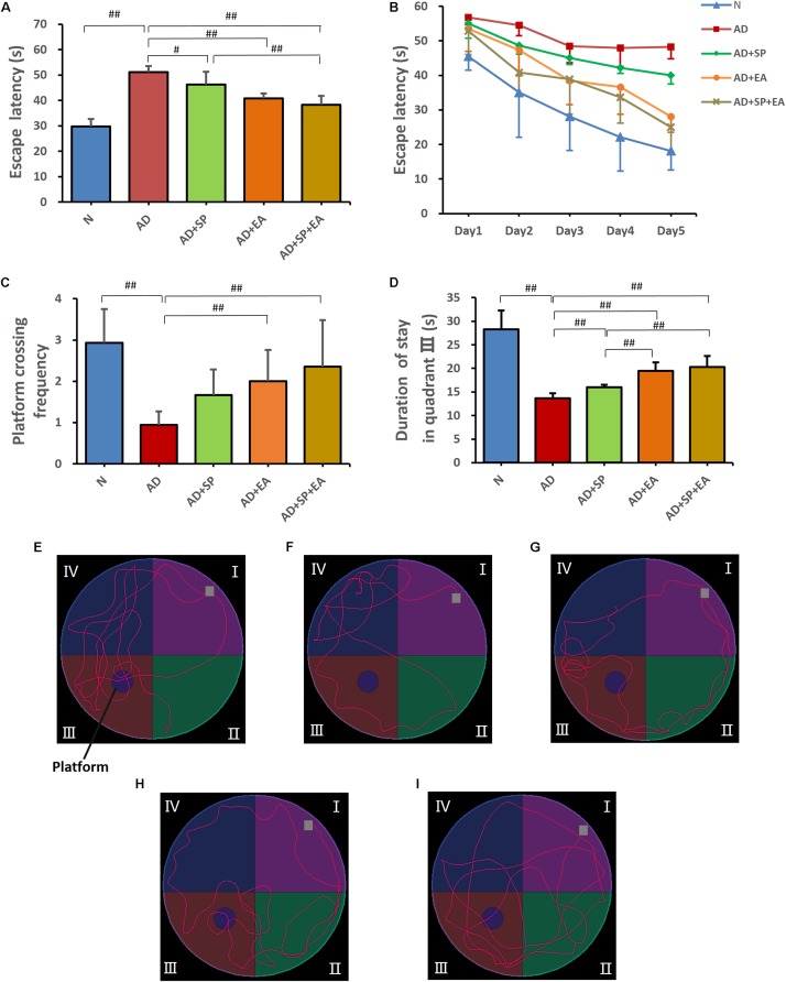
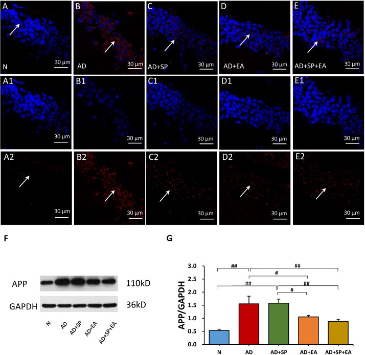
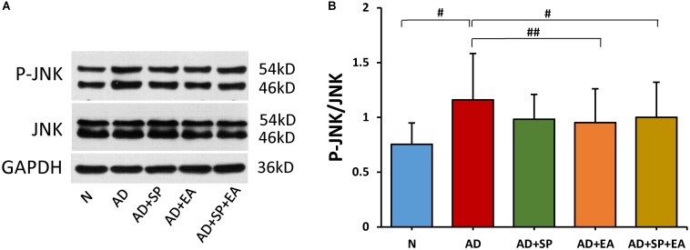
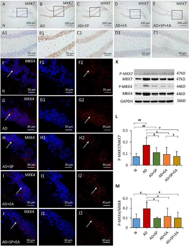
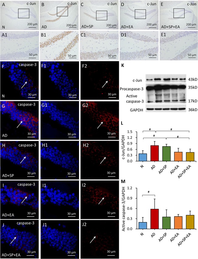
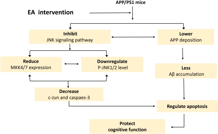
Electroacupuncture (EA) has become popular for its adjustable strength and frequency and easy quantification in the clinic and has demonstrated therapeutic potential for Alzheimer's disease (AD). However, the mechanism remains unknown. Abnormally activated c-Jun N-terminal kinase (JNK) has been closely related to the pathological process of AD. The aim of this study was to investigate the effect of EA on cognitive impairment and the role of the JNK signaling pathway in AD model amyloid precursor protein (APP)/presenilin 1 (PS1) mice. The memory and learning ability of each group was assessed using the Morris Water Maze (MWM). Immunofluorescence, immunohistochemistry and Western blot were performed to measure the expression of APP, JNK, phosphorylated (P-)JNK, mitogen-activated protein kinase 4 (MKK4), MKK7, c-Jun and caspase-3 in hippocampal tissue samples in APP/PS1 mice after EA intervention. Obvious cognitive deficits were observed in the AD model APP/PS1 mice in the MWM test and were associated with JNK signaling pathway activation and APP upregulation. Four weeks of EA significantly ameliorated the cognitive impairments and inhibited JNK signaling pathway activation and APP upregulation. Taken together, the findings demonstrated that EA can reverse cognitive deficits and substantially lower the burden of APP in AD model APP/PS1 mice, at least partially through inhibiting the JNK signaling pathway and regulating apoptosis signals. Therefore, EA may offer an effective alternative therapeutic approach for AD.

Keywords: APP/PS1 mice; Alzheimer’s disease; JNK signaling pathway; apoptosis; cognitive impairment; electroacupuncture.


Figures

FIGURE 1
FIGURE 1
Locations of acupoints we applied in this study. Red points indicate the three governor vessel acupoints respectively: GV20, GV29, and GV26. (A) Top views of GV20, GV29, and GV26. (B) Locations of GV20, GV29, and GV26.
FIGURE 2
FIGURE 2
Morris Water Maze test results after intervention (n = 10, mean ± SD). (A) Comparison of the average escape latency of all groups in training trials. (B) Trends of the escape latency of each group in training trials. (C) Platform crossing frequency of each group. (D) Duration of stay in quadrant III of each group. (E–I) Representative probe traces of each group. The water entry points are indicated by gray squares. n = 10 per group. #p < 0.05; ##p < 0.01.
FIGURE 3
FIGURE 3
Comparison of APP expression in the hippocampus of APP/PS1 mice in each group. (A–E) Comparison of APP expression using immunofluorescence. (A1–E2) Comparison of APP expression in the square frame of A-E in detail. APP: red; DAPI: blue; White arrows indicate positive expression of APP. (F,G) Comparison of APP expression using WB. Data are presented as the means ± SD, WB: n = 7 per group, immunofluorescence: n = 3 per group. #p < 0.05; ##p < 0.01.
FIGURE 4
FIGURE 4
Comparison of the expression of JNK in the hippocampus of APP/PS1 mice using WB. (A) The relative expression of hippocampal phosphorylated JNKs and total JNKs in APP/PS1 mice. (B) The ratio of p-JNK expression level to the total JNK level in the hippocampus of APP/PS1 mice. Data are presented as the means ± SD, WB: n = 7 per group. #p < 0.05; ##p < 0.01.
FIGURE 5
FIGURE 5
Comparison of MKK7 and MKK4 expression in the hippocampus of APP/PS1 mice in each group. (A–E) Comparison of MKK7 expression using immunohistochemistry. (A1–E1) Comparison of MKK7 expression in the square frame of (A–E) in detail. (F–F2,G–G2,H–H2,I–I2,J–J2) Comparison of MKK4 expression using immunofluorescence. MKK4: red; DAPI: blue; White arrows indicate positive expression of MKK4. (K) The relative expression of hippocampal phosphorylated MKK7 and MKK4 and total MKK7 and MKK4 in APP/PS1 mice. (L,M) The ratio of p-MKK7 and p-MKK4 expression level to the total MKK7 and MKK4 level in the hippocampus of APP/PS1 mice. Data are presented as the means ± SD, WB: n = 7 per group, immunofluorescence: n = 3 per group. #p < 0.05; ##p < 0.01.
FIGURE 6
FIGURE 6
Comparison of the expression of c-Jun and caspase-3 in the hippocampus of APP/PS1 mice in each group. (A–E) Comparison of c-Jun expression using immunohistochemistry. (A1–E1) Comparison of c-Jun expression in the square frame of (A–E) in detail. (F–F2,G–G2,H–H2,I–I2,J–J2) Comparison of caspase-3 expression using immunofluorescence. Caspase-3: red; DAPI: blue; White arrows indicate positive expression of MKK4. (K–M) Comparison of the expression of c-Jun and caspase-3 using WB. Data are presented as the means ± SD, WB: n = 7 per group, immunofluorescence: n = 3 per group. #p < 0.05.
FIGURE 7
FIGURE 7
Schematic of the mechanism by which EA intervention protects cognition in APP/PS1 mice. This schematic is derived from the results of this experiment combined with the results of our previous experiments.

References

    1. Bredesen D. E., Rao R. V., Mehlen P. (2006). Cell death in the nervous system. Nature 443 796–802. - PMC - PubMed
    1. Busquets O., Eritja A., Lopez B. M., Ettcheto M., Manzine P. R., Castro-Torres R. D., et al. (2019). Role of brain c-Jun N-terminal kinase 2 in the control of the insulin receptor and its relationship with cognitive performance in a high-fat diet pre-clinical model. J. Neurochem. 149 255–268. 10.1111/jnc.14682 - DOI - PubMed
    1. Cao J., Tang Y., Li Y., Gao K., Shi X., Li Z. (2017). Behavioral changes and hippocampus glucose metabolism in APP/PS1 transgenic mice via electro-acupuncture at governor vessel acupoints. Front. Aging Neurosci. 9:5. 10.3389/fnagi.2017.00005 - DOI - PMC - PubMed
    1. Chang J. L., Zhang H., Tan Z. J., Xiao J., Li S. R., Gao Y. (2017). Effect of electroacupuncture in patients with post-stroke motor aphasia neurolinguistic and neuroimaging characteristics. Wien. Klin. Wochenschr. 129 102–109. 10.1007/s00508-016-1070-1 - DOI - PubMed
    1. Chang Y. J., Linh N. H., Shih Y. H., Yu H. M., Li M. S., Chen Y. R. (2016). Alzheimer’s amyloid-beta sequesters caspase-3 in vitro via its C-terminal tail. ACS Chem. Neurosci. 7 1097–1106. 10.1021/acschemneuro.6b00049 - DOI - PubMed