. 2020 Feb 3:11:13.
doi: 10.3389/fphar.2020.00013. eCollection 2020.

MicroRNA-96 Promotes Vascular Repair in Oxygen-Induced Retinopathy-A Novel Uncovered Vasoprotective Function

Affiliations

MicroRNA-96 Promotes Vascular Repair in Oxygen-Induced Retinopathy-A Novel Uncovered Vasoprotective Function

Michel Desjarlais et al. Front Pharmacol. .

Abstract

Background and aims: Vascular degeneration is a hallmark in the pathogenesis of oxygen-induced retinopathy (OIR). Dysregulation of microRNAs (miRNAs), key regulators of genes expressions, has been implicated in the regulation of ocular angiogenesis. However, miRNAs specific functions in impaired vascular development during OIR are poorly understood. Herein, we identified miR-96 as one of the most highly expressed miRNAs in the retina and choroid during vascular development and investigated the potential role of miR-96 on microvascular degeneration in a rat OIR model.

Methods and results: Next generation sequencing (NGS) and qRT-PCR analysis showed that miR-96 maintain high levels of expression during ocular vascular development. Nevertheless, miR-96 was significantly downregulated in the retina and choroid of OIR rats (80% O2 from P5 to P10) during the phase of microvascular degeneration. Similarly, human retinal microvascular endothelial cells (HRMEC) subjected to hyperoxia (80% O2) showed a significant downregulation of miR-96 evaluated by qPCR. Interestingly, HRMEC supplemented with miR-96 regulated positively the expression of several key angiogenic factors including VEGF and ANG-2. To explore the angiogenic activity of miR-96 on HRMEC, we performed a gain/loss of function study. In a similar way to hyperoxia exposure, we observed a robust angiogenic impairment (tubulogenesis and migration) on HRMEC transfected with an antagomiR-96. Conversely, overexpression of miR-96 stimulated the angiogenic activity of HRMEC and protected against hyperoxia-induced endothelial dysfunction. Finally, we evaluated the potential vasoprotective function of miR-96 in OIR animals. Rat pups intravitreally supplemented with miR-96 mimic (1 mg/kg) displayed a significant preservation of retinal/choroidal microvessels at P10 compared to controls. This result was consistent with the maintenance of physiologic levels of VEGF and ANG-2 in the OIR retina.

Conclusion: This study demonstrates that miR-96 regulates the expression of angiogenic factors (VEGF/ANG-2) associated to the maintenance of retinal and choroidal microvasculature during physiological and pathological conditions. Intravitreal supplementation of miR-96 mimic could constitute a novel therapeutic strategy to improve vascular repair in OIR and other ischemic retinopathies.

Keywords: endothelial dysfunction; micro-RNA (miRNA); oxygen-induced retinopathy (OIR); vascular degeneration; vascular repair and angiogenesis.


Figures

Figure 1
Figure 1
miR-96 expression is downregulated in the retina and choroid of OIR rat and in human retinal endothelial cells subjected to hyperoxia. (A) NGS analyses showing miR-96 expression profile (RPM) in the retina and choroid during the vascular development of normoxic control rats vs OIR rats (OIR cycling-O2 model). (B, C) qRT-PCR validation analysis of miR-96 expression in the retina of normoxic control rats vs OIR rats (vasoobliteration: constant-O2 model) and respectively in Human Retinal Microvascular Endothelial Cell (HRMEC) subjected to hyperoxia (80% O2). Data are mean ± SEM. *P < 0.05 or **P < 0.01 vs CTL (control). N = 4–5/group.
Figure 2
Figure 2
miR-96 regulate simultaneous the expression level of VEGF/FGF-2/Ang2 in human retinal endothelial cells. To explore the regulatory function of miR-96 on HRMEC angiogenic factor production, cells were pre-transfected or not with 50 nM of miR-control (scrambled), miR-96 mimic or antagomiR-96 for 24 h, and subjected or not to hyperoxia (80% O2) for an additional 24 h. (A) qRT-PCR analyses of miR-96 transfection efficacy, and (B, C) screening of angiogenic factor expression modulated by miR-96. (D, E) qRT-PCR analyses of miR-96 protective effects in HMREC subjected to hyperoxia (D) and western blot validation and compiled histogram analysis of the regulatory role of miR-96 on VEGF, FGF-2 and Ang2 proteins expression levels. N = 3–4 experiments. Data are mean ± SEM. *P < 0.05 vs CTL (control) or # P < 0.05 vs hyperoxia.
Figure 3
Figure 3
Suppression of miR-96 leads to endothelial dysfunction, and miR-96 supplementation protects angiogenic properties of endothelial cells against hyperoxia. (A) In vitro evaluation of angiogenesis (tubulogenesis) using HRMEC cultured in Matrigel, and (B) cell migration scratch assay of HRMEC treated or not with a miR-control (scrambled), miR-96 mimic or antagomiR-96, and exposed or not to hyperoxia (80% O2). Data are mean ± SEM. *P < 0.05 vs CTL (control) or # P < 0.05 vs hyperoxia. N = 3–4 experiments.
Figure 4
Figure 4
Intraocular miR-96 (mimic) prevents retinal and choroidal vascular decay and facilitates revascularization in OIR model. OIR-subjected rats were injected intravitreally with a single dose (1 mg/kg) of miR-control or miR-96 mimic at P5 before hyperoxia exposure. Animals were then subjected to constant hyperoxia (80% O2) until P10. (A) qRT-PCR analysis showing miR-96 treatment efficacy in raising miR-96 expression. (B) Quantification analyze of total retinal vasoobliteration in OIR-subjected rats treated or not with miR-96 mimic. (C, D) Representative flatmount images of central and peripheral vascularization; histogram on right show compiled analysis. (E) Isolectin staining of sub-retina showing choroidal vessels. (F) Photographic image of choroid explant vascular sprouting in Matrigel after 5 days of incubation, in tissues from normoxia- and OIR-raised animals treated or not with miR-96. Data are mean ± SEM. **P < 0.01 or ***P < 0.001 vs CTL; #P < 0.05 or ##P < 0.01 or ###P < 0.001 vs OIR. N = 6–8/group.
Figure 5
Figure 5
miR-96 supplementation restores VEGF and Ang2 levels in retina of OIR-subjected rats. OIR rats where intravitreally injected with a single dose (1 mg/kg) of miR-CTL or miR-96 mimic at P5 before hyperoxia exposure. After treatments, the animals were subjected to constant hyperoxia (80% O2) until P10. (A, B, C) Respectively, qRT-PCR and western blot analysis of VEGF and Ang2 expression in the retina of OIR-subjected rats treated or not with miR-96 mimic. (D) Immunostaining of VEGF in retinal cross-section reveals its expression co-localized on endothelium (lectin-staining) in OIR-subjected rats treated or not with miR-96 mimic. *P <0.05 vs CTL or #vs OIR. N = 6–8 retinas/group.
Figure 6
Figure 6
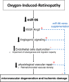
Schematic representation of vasoprotective function of miR-96 in OIR.

References

    1. Ayaz L., Dinc E. (2018). Evaluation of microRNA responses in ARPE-19 cells against the oxidative stress. Cutan Ocul. Toxicol. 37 (2), 121–126. 10.1080/15569527.2017.1355314 - DOI - PubMed
    1. Bai Y., Bai X., Wang Z., Zhang X., Ruan C., Miao J. (2011). MicroRNA-126 inhibits ischemia-induced retinal neovascularization via regulating angiogenic growth factors. Exp. Mol. Pathol. 91 (1), 471–477. 10.1016/j.yexmp.2011.04.016 - DOI - PubMed
    1. Bandara K. V., Michael M. Z., Gleadle J. M. (2017). MicroRNA biogenesis in hypoxia. Microrna 6 (2), 80–96. 10.2174/2211536606666170313114821 - DOI - PubMed
    1. Banerjee J., Khanna S., Bhattacharya A. (2017). MicroRNA regulation of oxidative stress. Oxid. Med. Cell Longev. 2017, 2872156. 10.1155/2017/2872156 - DOI - PMC - PubMed
    1. Battaglia R., Palini S., Vento M. E., La Ferlita A., Lo Faro M. J., Caroppo E., et al. (2019). Identification of extracellular vesicles and characterization of miRNA expression profiles in human blastocoel fluid. Sci. Rep. 9 (1), 84. 10.1038/s41598-018-36452-7 - DOI - PMC - PubMed