Review
. 2020 Feb 5:11:56.
doi: 10.3389/fphys.2020.00056. eCollection 2020.

Calcification in Atherosclerotic Plaque Vulnerability: Friend or Foe?

Affiliations
Review

Calcification in Atherosclerotic Plaque Vulnerability: Friend or Foe?

Xuan Shi et al. Front Physiol. .

Abstract

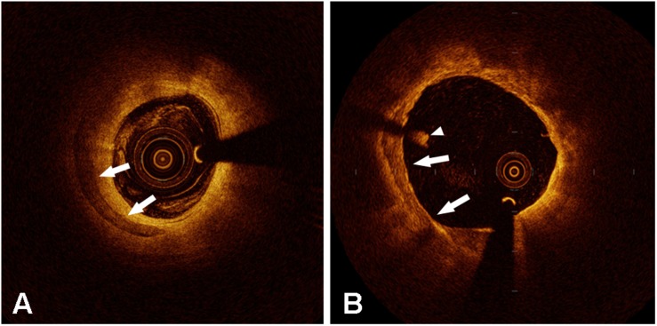
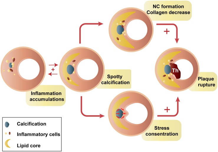
Calcification is a clinical marker of atherosclerosis. This review focuses on recent findings on the association between calcification and plaque vulnerability. Calcified plaques have traditionally been regarded as stable atheromas, those causing stenosis may be more stable than non-calcified plaques. With the advances in intravascular imaging technology, the detection of the calcification and its surrounding plaque components have evolved. Microcalcifications and spotty calcifications represent an active stage of vascular calcification correlated with inflammation, whereas the degree of plaque calcification is strongly inversely related to macrophage infiltration. Asymptomatic patients have a higher content of plaque calcification than that in symptomatic patients. The effect of calcification might be biphasic. Plaque rupture has been shown to correlate positively with the number of spotty calcifications, and inversely with the number of large calcifications. There may be certain stages of calcium deposition that may be more atherogenic. Moreover, superficial calcifications are independently associated with plaque rupture and intraplaque hemorrhage, which may be due to the concentrated and asymmetrical distribution of biological stress in plaques. Conclusively, calcification of differential amounts, sizes, shapes, and positions may play differential roles in plaque homeostasis. The surrounding environments around the calcification within plaques also have impacts on plaque homeostasis. The interactive effects of these important factors of calcifications and plaques still await further study.

Keywords: atherosclerosis; calcification; inflammation; optical coherence tomography; pathology; plaque.


Figures

FIGURE 1
FIGURE 1
Representative optical coherence tomography images of calcification (arrow in panels A and B). The arrowhead shows an intraluminal white thrombus.
FIGURE 2
FIGURE 2
Hypothesis of spotty calcification in plaque rupture. Two main ways that spotty calcifications lead to plaque rupture: inflammatory cytokines from macrophages activate osteogenic differentiation, contributing to early stages of calcification. Then, a positive feedback loop between inflammation and calcification produces spotty calcification, stimulating accelerated plaque progression, including greater inflammatory burden, a larger necrotic core, and less collagen (Aikawa et al., 2007; Kataoka et al., 2012; Pu et al., 2014, 2016a; Hsu et al., 2016). In contrast, calcification neighboring the lipid pool, especially that near the fibrous cap, may intensify failure stress and cause plaque rupture (Bluestein et al., 2008; Hoshino et al., 2009). NC, necrotic core; Th, thrombus.

References

    1. Abedin M., Tintut Y., Demer L. L. (2004). Vascular calcification: mechanisms and clinical ramifications. Arterioscler. Thromb. Vasc. Biol. 24 1161–1170. 10.1161/01.ATV.0000133194.94939.42 - DOI - PubMed
    1. Afolabi A., Mustafina I., Zhao L., Li L., Sun R., Hu S., et al. (2018). Does spotty calcification attenuate the response of nonculprit plaque to statin therapy?: a serial optical coherence tomography study. Catheter. Cardiovasc. Interv. 91 582–590. 10.1002/ccd.27496 - DOI - PubMed
    1. Aikawa E., Nahrendorf M., Figueiredo J. L., Swirski F. K., Shtatland T., Kohler R. H., et al. (2007). Osteogenesis associates with inflammation in early-stage atherosclerosis evaluated by molecular imaging in vivo. Circulation 116 2841–2850. 10.1161/CIRCULATIONAHA.107.732867 - DOI - PubMed
    1. Amizuka N., Hasegawa T., Oda K., Luiz de Freitas P. H., Hoshi K., Li M., et al. (2012). Histology of epiphyseal cartilage calcification and endochondral ossification. Front. Biosci. 4 2085–2100. 10.2741/526 - DOI - PubMed
    1. Bluestein D., Alemu Y., Avrahami I., Gharib M., Dumont K., Ricotta J. J., et al. (2008). Influence of microcalcifications on vulnerable plaque mechanics using FSI modeling. J. Biomech. 41 1111–1118. 10.1016/j.jbiomech.2007.11.029 - DOI - PubMed

LinkOut - more resources