Review
. 2020 Feb 14:11:34.
doi: 10.3389/fneur.2020.00034. eCollection 2020.

Posterior Reversible Encephalopathy Syndrome and Reversible Cerebral Vasoconstriction Syndrome: Clinical and Radiological Considerations

Affiliations
Review

Posterior Reversible Encephalopathy Syndrome and Reversible Cerebral Vasoconstriction Syndrome: Clinical and Radiological Considerations

Fabio Pilato et al. Front Neurol. .

Abstract

Posterior reversible encephalopathy syndrome (PRES) and reversible cerebral vasoconstriction syndrome (RCVS) are relatively uncommon neurological disorders, but their detection has been increasing mainly due to clinical awareness and spreading of magnetic resonance imaging (MRI). Because these syndromes share some common clinical and radiologic features and occasionally occur in the same patient, misdiagnosis may occur. PRES is characterized by varied neurological symptoms including headache, impaired visual acuity or visual field deficit, confusion, disorders of consciousness, seizures, and motor deficits often associated to peculiar neuroradiological pattern even if uncommon localization and ischemic or hemorrhagic lesions were described. RCVS is a group of diseases typically associated with severe headaches and reversible segmental vasoconstriction of cerebral arteries, often complicated by ischemic or hemorrhagic stroke. Pathophysiological basis of PRES and RCVS are still debated but, because they share some risk factors and clinical features, a possible common origin has been supposed. Clinical course is usually self-limiting, but prognosis may fluctuate from complete recovery to death due to complications of ischemic stroke or intracranial hemorrhage. Neuroradiological techniques such as digital angiography and MRI are helpful in the diagnostic pathway and a possible prognostic role of MRI has been suggested. This review will serve to summarize clinical, neuroradiological features and controversies underlying both syndromes that may mislead the diagnostic pathway and their possible relationship with pathophysiology, clinical course, and prognosis.

Keywords: PRES; RCVS; call-fleming syndrome; magnetic resonance imaging; posterior reversible encephalopathy syndrome; reversible cerebral vasoconstriction syndrome; reversible posterior leukoencephalopathy syndrome.


Figures

Figure 1
Figure 1
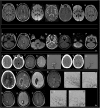
(a) Typical dominant parietal–occipital pattern in a patient with PRES at the onset (A–E) and after 15 days (F,G). (A,B,G) FLAIR MR images; (C,F) DWI MR images; (D) ADC map; (E) T1 C+ MR image. Edema involves the parietal and occipital cortex and white matter (A,B); small, patchy, or punctate hyperintensity in DWI (white arrows in C) corresponding to hypointensity in ADC map (white arrows in D) characterize the cytotoxic edema within diffuse vasogenic edema; gyral or leptomeningeal enhancement is shown in occipital regions (E). Note resolution of the lesions 15 days after the onset (F,G). Atypical involvement of the brainstem associated to occipital pattern in a patient with PRES at the onset (H–N) and after 18 days (O,P). (H,I,P) FLAIR MR images; (L,O) DWI MR image; (M) ADC map; (N) T1 C+ MR image. Edema involves the right cerebellum, brainstem, and occipital cortex and white matter (H,I); iso-intensity with punctate foci of hyperintensity in DWI (white arrows in L) and hyperintense signal in ADC characterizes the vasogenic edema (white arrow in M), no enhancement is shown (N). Note resolution of the lesions 18 days after the onset (O,P). (b) Intracranial subarachnoid hemorrhage in a patient with RCVS at the onset (A–E) and after 2 months (F–H). Axial CT (A,B) shows hyperdense subarachnoid hemorrhage in the right frontal (white arrow in A) and left occipital lobes (white arrow in B); axial FLAIR MR (C) confirms subarachnoid hemorrhage as hyperintense sulci (white arrow in C); SWI MR images (D) show a component of subarachnoid hemorrhage as hypointense focus within a frontal sulcus (white arrow in D); catheter angiography of vertebro-basilar arteries demonstrate vessel irregularities with focal vasoconstriction (white arrow in E). Note resolution of SAH (F,G) and vessel irregularities (H) after 2 months. Intraparenchymal hematoma and subarachnoid hemorrhage in a patient with post-partum RCVS at the onset (I–P) and after 3 months (Q–U). Axial CT (I) shows hyperdense parenchymal hematoma in the right frontal lobe (white arrow in I) and subarachnoid hemorrhage in the left frontal lobe (white head of arrows in I); FLAIR MR (L) shows vasogenic edema marginally at the right parenchymal hematoma (white arrow in L) and left subarachnoid hemorrhage as hyperintense sulci (white head of arrows in L); SWI MR image (M) shows the hypointense signal of the parenchymal and subarachnoid hemorrhage due to acute phase of hemorrhage; contrast-enhanced MRA (CE-MRA) (N) images show vasoconstriction of some distal branches of middle cerebral arteries (with arrows in N); catheter angiography of internal carotids confirms diffuse irregularities with multifocal narrowings throughout the cerebral vasculature with a “string-of-beads” appearance (white arrows in O,P). Note reduction of ICH and SAH (Q–S) and disappearance of multifocal narrowings of distal branches of middle cerebral arteries after 3 months (T,U).

Similar articles

Cited by

References

    1. Hinchey J, Chaves C, Appignani B, Breen J, Pao L, Wang A, et al. . A reversible posterior leukoencephalopathy syndrome. N Engl J Med. (1996) 334:494–500. 10.1056/NEJM199602223340803 - DOI - PubMed
    1. Ducros A. Reversible cerebral vasoconstriction syndrome. Lancet Neurol. (2012) 11:906–17. 10.1016/S1474-4422(12)70135-7 - DOI - PubMed
    1. Miller TR, Shivashankar R, Mossa-Basha M, Gandhi D. Reversible cerebral vasoconstriction syndrome, part 1: epidemiology, pathogenesis, and clinical course. AJNR Am J Neuroradiol. (2015) 36:1392–9. 10.3174/ajnr.A4214 - DOI - PMC - PubMed
    1. Fugate JE, Rabinstein AA. Posterior reversible encephalopathy syndrome: clinical and radiological manifestations, pathophysiology, and outstanding questions. Lancet Neurol. (2015) 14:914–25. 10.1016/s1474-4422(15)00111-8 - DOI - PubMed
    1. Covarrubias DJ, Luetmer PH, Campeau NG. Posterior reversible encephalopathy syndrome: prognostic utility of quantitative diffusion-weighted MR images. AJNR Am J Neuroradiol. (2002) 23:1038–48. - PMC - PubMed