. 2020 Jan 29:11:65.
doi: 10.3389/fmicb.2020.00065. eCollection 2020.

Finegoldia magna, an Anaerobic Gram-Positive Bacterium of the Normal Human Microbiota, Induces Inflammation by Activating Neutrophils

Affiliations

Finegoldia magna, an Anaerobic Gram-Positive Bacterium of the Normal Human Microbiota, Induces Inflammation by Activating Neutrophils

Ariane Neumann et al. Front Microbiol. .

Abstract

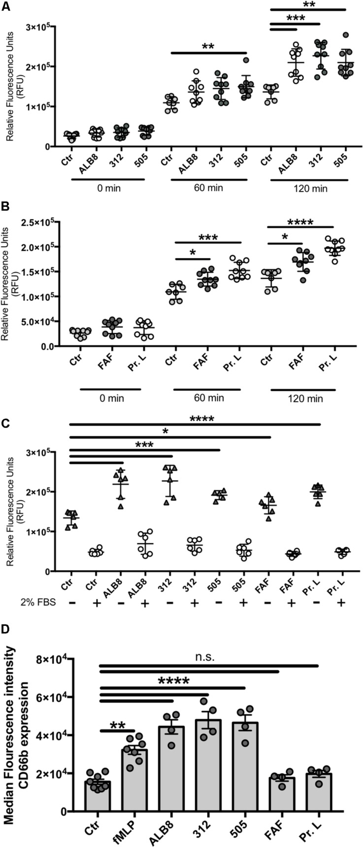
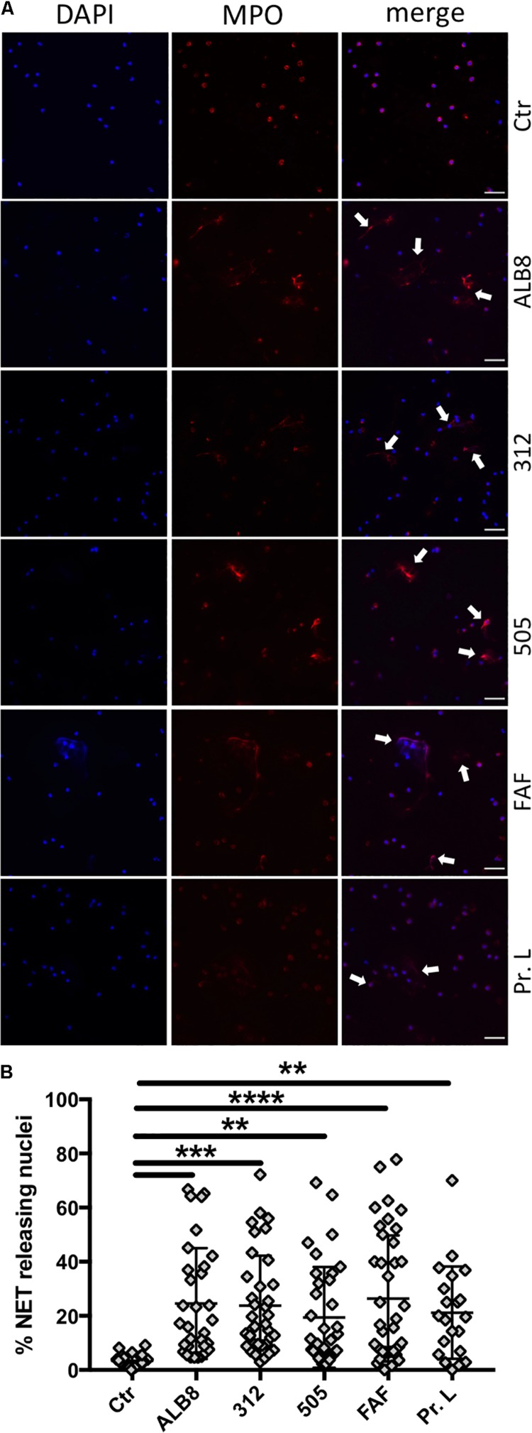
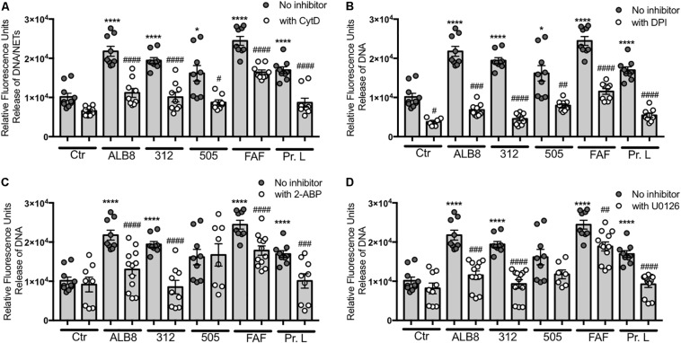
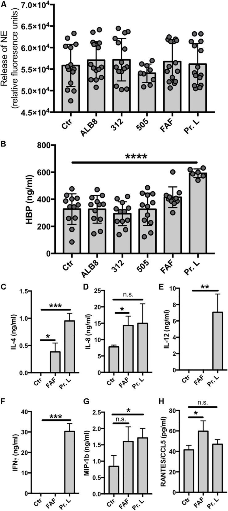
The Gram-positive anaerobic commensal Finegoldia magna colonizes the skin and other non-sterile body surfaces, and is an important opportunistic pathogen. Here we analyzed the effect of F. magna on human primary neutrophils. F. magna strains ALB8 (expressing protein FAF), 312 (expressing protein L) and 505 (naturally lacking both protein FAF and L) as well as their associated proteins activate neutrophils to release reactive oxygen species, an indication for neutrophil oxidative burst. Co-incubation of neutrophils with the bacteria leads to a strong increase of CD66b surface expression, another indicator for neutrophil activation. Furthermore, all tested stimuli triggered the release of NETs from the activated neutrophils, pointing to a host defense mechanism in response to the tested stimuli. This phenotype is dependent on actin rearrangement, NADPH oxidases and the ERK1/2 pathway. Proteins FAF and L also induced the secretion of several pro-inflammatory neutrophil proteins; HBP, IL-8 and INFγ. This study shows for the first time a direct interaction of F. magna with human neutrophils and suggests that the activation of neutrophils plays a role in F. magna pathogenesis.

Keywords: CD66b expression; GPAC; NETs; anaerobic Gram-positive cocci; host-pathogen interactions; inflammation; neutrophils.


Figures

FIGURE 1
FIGURE 1
Finegoldia magna bacteria and respective proteins activate neutrophils. Neutrophils were incubated with (A) heat inactivated F. magna strains ALB8, 312 or 505 or with (B) purified protein FAF and protein L. Production of ROS was measured every 60 min and is displayed as relative fluorescence units (RFU). (C) ROS production was measured in presence and absence of 2% FBS, which reduced all stimuli-triggered ROS production. Control (Ctr) represents unstimulated neutrophils. (D) Neutrophils incubated as above were analyzed by flow cytometry. The median fluorescence intensity indicates the CD66b surface expression on neutrophils in response to F. magna strains and purified proteins FAF and protein L. All data represent mean ± SEM of 3 independent experiments. *p ≤ 0.05, **p ≤ 0.01, ***p ≤ 0.001, ****p ≤ 0.0001.
FIGURE 2
FIGURE 2
F. magna bacteria and proteins induce NET formation. (A) Neutrophils were incubated with heat inactivated F. magna strains ALB8, 312 or 505 or with purified proteins FAF and L. Control (Ctr) represents unstimulated neutrophils. NETs (white arrows) were visualized with DAPI (blue) and MPO (red), scale bar is 50 μm. (B) NET-releasing nuclei were quantified using ImageJ software and are displayed as percentage. Data represent mean ± SEM of 4 independent experiments. **p ≤ 0.01, ***p ≤ 0.001, ****p ≤ 0.0001.
FIGURE 3
FIGURE 3
Actin rearrangement and NADPH oxidases are involved in F. magna-mediated NET release. Neutrophils were co-incubated with heat inactivated F. magna bacteria or proteins FAF and L for 2 h. The release of NETs was blocked by the addition of (A) 20 μM Cytochalasin D (CytD), (B) 10 μM DPI, (C) 50 μM 2-ABP, (D) 50 μM U0126. All data represent mean ± SEM of 3–5 independent experiments. *p ≤ 0.05, ****p ≤ 0.0001 to Ctr; #p ≤ 0.05, ##p ≤ 0.01, ###p ≤ 0.001, ####p ≤ 0.0001 to stimuli without inhibitor.
FIGURE 4
FIGURE 4
Protein L triggers HBP and cytokine release. (A–H) Neutrophils were incubated with F. magna bacteria or their proteins for 2 h and supernatants were collected for (A) NE enzyme activity, (B) HPB release or (C–H) cytokine profiling. (A) Enzyme activity was measured using NE EnzChek assay kit. None of the used stimuli triggered enzyme activity. (B) Protein L significantly induced the release of HBP. Whole bacteria and protein F had no effect on the HBP release. (C–H) Cytokine profile was determined after co-incubation of neutrophils with F. magna-derived proteins FAF (3.8 μg/ml) or protein L (3 μg/ml) for 2 h. (C) IL-4, (D) IL-8, (E) IL-12, F) INFγ, (G) MIP-1b and (H) RANTES/CCL5. Data represent mean ± SEM of 3 independent experiments. *p ≤ 0.05, **p ≤ 0.01, ***p ≤ 0.001, ****p ≤ 0.0001.

References

    1. Akerstrom B., Bjorck L. (1989). Protein L: an immunoglobulin light chain-binding bacterial protein. Characterization of binding and physicochemical properties. J. Biol. Chem. 264 19740–19746. - PubMed
    1. Akerstrom B., Bjorck L. (2009). Bacterial surface protein L binds and inactivates neutrophil proteins S100A8/A9. J. Immunol. 183 4583–4592. 10.4049/jimmunol.0901487 - DOI - PubMed
    1. Anderson A. L., Zheng Y., Song D., Larosa D., Van Rooijen N., Kierstein G., et al. (2012). The B-cell superantigen Finegoldia magna protein L causes pulmonary inflammation by a mechanism dependent on MyD88 but not B cells or immunoglobulins. Inflamm. Res. 61 161–169. 10.1007/s00011-012-0436-8 - DOI - PMC - PubMed
    1. Berge A., Bjorck L. (1995). Streptococcal cysteine proteinase releases biologically active fragments of streptococcal surface proteins. J. Biol. Chem. 270 9862–9867. 10.1074/jbc.270.17.9862 - DOI - PubMed
    1. Bjorck L. (1988). Protein L. A novel bacterial cell wall protein with affinity for Ig L chains. J. Immunol. 140 1194–1197. - PubMed