. 2020 Feb 13:11:114.
doi: 10.3389/fmicb.2020.00114. eCollection 2020.

A Novel Polyester Hydrolase From the Marine Bacterium Pseudomonas aestusnigri - Structural and Functional Insights

Affiliations

A Novel Polyester Hydrolase From the Marine Bacterium Pseudomonas aestusnigri - Structural and Functional Insights

Alexander Bollinger et al. Front Microbiol. .

Abstract

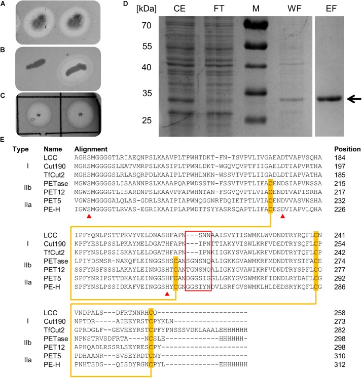
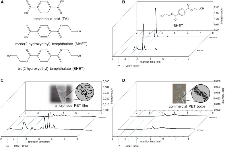
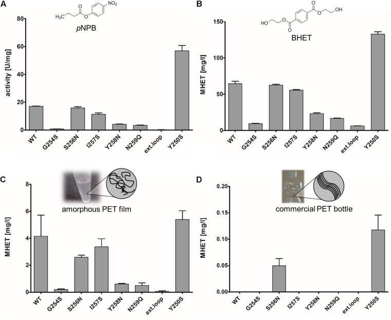
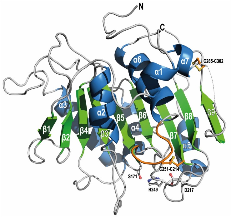
Biodegradation of synthetic polymers, in particular polyethylene terephthalate (PET), is of great importance, since environmental pollution with PET and other plastics has become a severe global problem. Here, we report on the polyester degrading ability of a novel carboxylic ester hydrolase identified in the genome of the marine hydrocarbonoclastic bacterium Pseudomonas aestusnigri VGXO14 T . The enzyme, designated PE-H, belongs to the type IIa family of PET hydrolytic enzymes as indicated by amino acid sequence homology. It was produced in Escherichia coli, purified and its crystal structure was solved at 1.09 Å resolution representing the first structure of a type IIa PET hydrolytic enzyme. The structure shows a typical α/β-hydrolase fold and high structural homology to known polyester hydrolases. PET hydrolysis was detected at 30°C with amorphous PET film (PETa), but not with PET film from a commercial PET bottle (PETb). A rational mutagenesis study to improve the PET degrading potential of PE-H yielded variant PE-H (Y250S) which showed improved activity, ultimately also allowing the hydrolysis of PETb. The crystal structure of this variant solved at 1.35 Å resolution allowed to rationalize the improvement of enzymatic activity. A PET oligomer binding model was proposed by molecular docking computations. Our results indicate a significant potential of the marine bacterium P. aestusnigri for PET degradation.

Keywords: PET; Pseudomonas aestusnigri; crystal structure; marine bacteria; polyester degradation; polyethylene terephthalate.


Figures

FIGURE 1
FIGURE 1
(A) Colonies of P. aestusnigri grown on an Impranil DLN containing agar plate show clearing halos, indicating polyester hydrolase activity. (B) Hydrolytic activity of E. coli BL21(DE3) empty vector control (left) and PE-H production strain (right) on Impranil DLN containing agar plate. (C) Hydrolytic activity of purified PE-H on Impranil DLN containing agar plate. (D) Coomassie brilliant blue stained gel after SDS-PAGE of cell extracts of E. coli BL21(DE3) containing plasmid pET22b_PE-Hc6H before and after purification by IMAC; the position of PE-H is indicated by an arrow. Lanes contained cell extract (CE), flow through (FT), washing step (WF), and eluted protein (EF); the size of the molecular weight standards (M) are indicated on the left. (E) Part of a multiple sequence alignment of PE-H with amino acid sequences of different PET hydrolytic enzymes using the program Clustal Omega. The full length alignment can be found in the Supplementary Figure S1. The enzymes were assigned to different types of polyester hydrolases (Joo et al., 2018). Amino acid residues of the catalytic triad are marked by a red triangle, disulfide forming cysteine residues are highlighted in orange and connected by an orange line. Amino acids forming the extended loop region which is specific for type II PET hydrolytic enzymes are framed in red. Abbreviations are: leaf-branch compost metagenome cutinase (LCC); Saccharomonospora viridis cutinase (Cut190); Thermobifida fusca cutinase (TfCut2); Ideonella sakaiensis PET hydrolase (PETase); Polyangium brachysporum PET hydrolase (PET12); Oleispira antarctica PET hydrolase (PET5).
FIGURE 2
FIGURE 2
Hydrolysis of PET by PE-H. (A) Structural formula of aromatic reaction products obtained by hydrolysis of PET or BHET. (B) UPLC analysis of reaction products obtained with PE-H and the substrates bis(2-hydroxyethyl) terephthalate (BHET), (C) amorphous PET film (PETa) and (D) PET film derived from a commercial single use PET bottle (PETb). The UPLC trace at 240 nm wavelength is shown for the control reaction without addition of the enzyme (trace in the back, labeled as control), and the enzyme reaction (trace in front, labeled PE-H). The retention times of the standard compounds terephthalic acid (TA), and bis(2-hydroxyethyl) terephthalate (BHET) as well as of the reaction product mono(2-hydroxyethyl) terephthalate (MHET) are indicated by blue dashed lines. The respective substrate of the reaction is depicted in the back of each diagram. For BHET, the structural formula is given; for PET films, a cartoon representation is shown to illustrate the different arrangement of PET fibers in amorphous and crystalline films.
FIGURE 3
FIGURE 3
Enzymatic activity of PE-H and different variants constructed by site directed mutagenesis. Substrates were (A) 4-nitrophenyl butyrate (pNPB), (B) bis(2-hydroxyethyl) terephthalate (BHET), (C) amorphous PET film, and (D) PET from a commercial single use bottle. Standard deviations of three individual reactions are shown as error bars. The respective substrate is depicted above each diagram. For pNPB and BHET, structural formulas are shown; for amorphous and commercial PET a cartoon representation is shown to illustrate the different arrangement of PET fibers.
FIGURE 4
FIGURE 4
Overall fold of WT PE-H (PDB code 6SBN) in cartoon representation. The short stretch (aa 286–291) is not visible (gray dashed line). The α/β-fold consists of a central twisted β-sheet composed of 9 β-strands flanked by 7 α-helices on both sides. The extended loop region and the Cys residues (ball-and-sticks) forming the disulfide bonds are highlighted in orange. Residues building the catalytic triad are shown as gray ball-and-sticks with labels. Rotations between the structure depictions are indicated in Supplementary Figure S4.
FIGURE 5
FIGURE 5
Superposition of PE-H (PDB code 6SBN, silver) and its variant Y250S (PDB code 6SCD, wheat). (A) The top view on the active site and the different orientation of the two loops is shown. Residues of the catalytic triad are depicted as sticks with labels, loops of PE-H are colored in green to highlight the structural differences. (B) A zoom in the active site cleft with the catalytic residues depicted as lines. Remarkably, Y250 (gray sticks) makes a polar contact with E102 (green sticks) in the wild type protein, the distance is shown as a dashed line and given in Å, whereas in variant Y250S, residue E102 (wheat sticks) is far apart [color code as in (A)]. (C) Surface representation of WT PE-H (silver) and variant Y250S (wheat), on top, both structures are superposed with the catalytic cleft indicated by a dashed circle. In the panels, the differences in both structures are displayed in detail; with WT PE-H in the left panel and variant Y250S in the right panel. Residues confining the active site cleft are depicted as sticks, labeled, and connected by dotted lines, with the corresponding distances given in Å. Rotations between the different structure depictions are indicated in Supplementary Figure S4.
FIGURE 6
FIGURE 6
Overlay of PE-H WT (PDB code 6SBN, gray) with (A) Cut190 (PDB code 4WFI, green) and (B) PETase (PDB code 6ANE, orange). Residues of the active site and disulfide bonds are represented as sticks with labels (according to PE-H numbering). (C) Superimposition of PE-H (PDB code 6SBN, gray), PETase (PDB code 6ANE, orange), and Cut190 (PDB code 4WFI, green). Five regions of the superimposition are represented in full tone on fade-out color to highlight the differences of the structures. In (D) the structures are turned from bottom to top.
FIGURE 7
FIGURE 7
Predicted ligand binding modes in wild type PE-H and variant Y250S. The predicted binding poses of BHET (magenta), MHET (yellow), and 2-HE(MHET)4 (brown) in WT PE-H (navy) and the variant Y250S (cyan). In (A,C,F) S171 is shown in orange, and in (B,D,E) the catalytic triad (S171, D217, and H249) is shown in orange and interacting residues are shown in white. (A) In wild type PE-H, BHET and MHET bind to a groove adjacent to the catalytic site (white arrow). (B) BHET and MHET bind to the hydrophobic groove and are stabilized by hydrogen bonding interactions with S103, D106, S248, and S256. 2-HE(MHET)4 binds similarly to BHET and MHET in the groove adjacent to the catalytic site. (C) In the variant Y250S, MHET binds to the catalytic site, while BHET occupies the hydrophobic groove. (D) MHET binds to the catalytic site and is stabilized by hydrophobic interactions to F98, V99, M172, and I219 such that S171 can attack the carbonyl carbon for ester hydrolysis. (E) A second binding pose of BHET binds similar to MHET. (F) Proposed mechanism of PET polymer interaction. Residues G254, Y258, and N259, which when substituted decrease esterase activity, are shown in red. One polymer unit (stylized green rectangle) binds to the groove adjacent to the catalytic site, a second unit bridges the distance to the catalytic site, and a third unit is cleaved from the polymer chain at the catalytic unit.

References

    1. Adrados A., de Marco I., Caballero B. M., López A., Laresgoiti M. F., Torres A. (2012). Pyrolysis of plastic packaging waste: a comparison of plastic residuals from material recovery facilities with simulated plastic waste. Waste Manag. 32 826–832. 10.1016/j.wasman.2011.06.016 - DOI - PubMed
    1. Almagro Armenteros J. J., Tsirigos K. D., Sønderby C. K., Petersen T. N., Winther O., Brunak S., et al. (2019). SignalP 5.0 improves signal peptide predictions using deep neural networks. Nat. Biotechnol. 37 420–423. 10.1038/s41587-019-0036-z - DOI - PubMed
    1. Altschul S. F., Gish W., Miller W., Myers E. W., Lipman D. J. (1990). Basic local alignment search tool. J. Mol. Biol. 215 403–410. 10.1016/S0022-2836(05)80360-2 - DOI - PubMed
    1. Austin H. P., Allen M. D., Donohoe B. S., Rorrer N. A., Kearns F. L., Silveira R. L., et al. (2018). Characterization and engineering of a plastic-degrading aromatic polyesterase. Proc. Natl. Acad. Sci. U.S.A. 115 E4350–E4357. 10.1073/pnas.1718804115 - DOI - PMC - PubMed
    1. Barth M., Oeser T., Wei R., Then J., Schmidt J., Zimmermann W. (2015). Effect of hydrolysis products on the enzymatic degradation of polyethylene terephthalate nanoparticles by a polyester hydrolase from Thermobifida fusca. Biochem. Eng. J. 93 222–228. 10.1016/J.BEJ.2014.10.012 - DOI

LinkOut - more resources