Review
. 2020 Jan 24:11:26.
doi: 10.3389/fimmu.2020.00026. eCollection 2020.

Novel Insights Into Immune Systems of Bats

Affiliations
Review

Novel Insights Into Immune Systems of Bats

Arinjay Banerjee et al. Front Immunol. .

Abstract

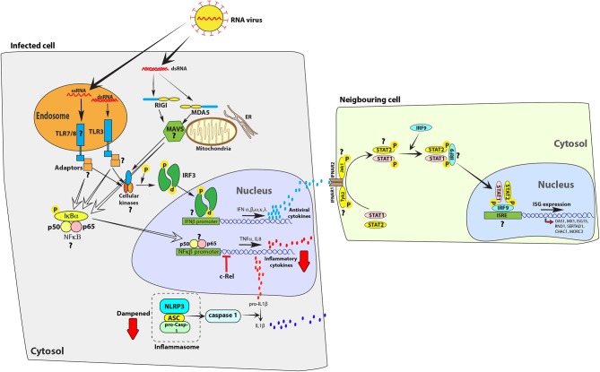
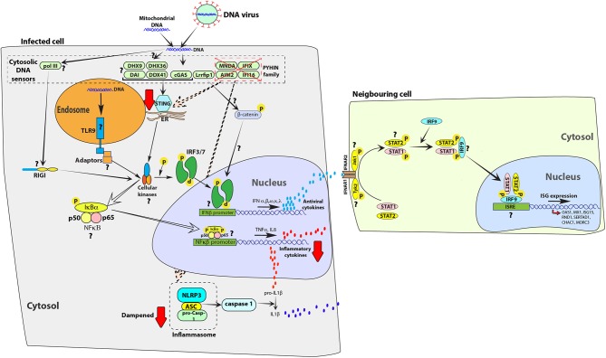
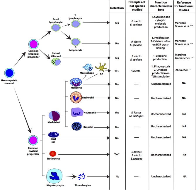
In recent years, viruses similar to those that cause serious disease in humans and other mammals have been detected in apparently healthy bats. These include filoviruses, paramyxoviruses, and coronaviruses that cause severe diseases such as Ebola virus disease, Marburg haemorrhagic fever and severe acute respiratory syndrome (SARS) in humans. The evolution of flight in bats seem to have selected for a unique set of antiviral immune responses that control virus propagation, while limiting self-damaging inflammatory responses. Here, we summarize our current understanding of antiviral immune responses in bats and discuss their ability to co-exist with emerging viruses that cause serious disease in other mammals. We highlight how this knowledge may help us to predict viral spillovers into new hosts and discuss future directions for the field.

Keywords: antiviral; bats (Chiroptera); emerging viruses; innate and adaptive immune response; interferon; virus.


Figures

Figure 1
Figure 1
Bat cells mount an antiviral response to RNA viruses, but limit the expression of inflammatory cytokines. Infection with RNA viruses, such as Sendai virus, or transfecting cells with surrogate double-stranded RNA [poly(I:C)] or single-stranded RNA is detected by Toll-like receptors (TLRs) 3, 7 and 8 or cytosolic receptors retinoic acid-inducible gene-I (RIG-I) and melanoma differentiation-associated protein 5 (MDA5). Activation of these receptors activate downstream adaptor proteins, such as mitochondrial antiviral signaling protein (MAVS). Adaptor proteins activate cellular kinases, such as TANK-binding kinase 1 protein (TBK1), which in-turn activate interferon regulatory factor 3 (IRF3) or IRF7 and nuclear factor kappa-light-chain-enhancer of activated B cells (NFκB) to stimulate the expression of interferons (IFNs), such as IFNs α, β, ω, κ, and λ, and pro-inflammatory cytokines, such as IL8, TNFα, and IL1β, respectively. IFNs bind to interferon α/β receptors (IFNAR; IFNAR1 and IFNAR2) on infected and neighboring cells to activate the JAK-STAT signaling pathway via kinases such as Janus kinase 1 (Jak1) and tyrosine kinase 2 (Tyk2) that phosphorylate signal transducer and activator of transcription (STAT) proteins. Phosphorylated STAT proteins (STAT1 and STAT2) merge with IRF9 and induce the expression of interferon stimulated genes (ISGs), such as OAS1 and Mx1. However, unlike in human cells, the parallel activation of pro-inflammatory cytokines is dampened in bat cells. c-Rel, a protein from the NFκB family of proteins binds to the TNFα promoter to inhibit activation of this pro-inflammatory cytokine in E. fuscus cells (49). The bat NLRP3 inflammasome activation is dampened, reducing the ability of bat cells to produce IL1β, a key inflammatory cytokine (17). In the figure, red arrows indicate a dampened response in the pathway, relative to human cells. Question marks (?) highlight pathways and molecular homologs that have not been characterized or identified in bats. The data have been compiled from studies in different species and one finding may not represent a universal bat response. ER, endoplasmic reticulum.
Figure 2
Figure 2
Exogenous and self-DNA sensing pathways are dampened in bat cells. In human cells, endosomal TLR9 (35) and cytosolic receptors from the PYHIN family (86) [absent in melanoma 2 (AIM2), IFN-inducible gene 16 (IFI16), myeloid cell nuclear differentiation antigen (MNDA) and IFN-inducible protein X (IFIX)], cyclic GMP-AMP synthase (cGAS) (87), DNA-dependent activator of IFN-regulatory factor (DAI) (88), RNA polymerase III (PolIII) (89), LRR binding FLII interacting protein 1 (Lrrfip1) (90), DDX41 (91), DExH-Box helicase 9 (DHX9) and DEAH-Box helicase 36 (DHX36) (92) can detect exogenous and self-DNA (86, 93). On binding to DNA, these receptors signal through adaptor proteins to activate cellular kinases, such as TANK-binding kinase 1 protein (TBK1), which in-turn activates transcription factors, such as interferon regulatory factor 3 (IRF3) or IRF7 and nuclear factor kappa-light-chain-enhancer of activated B cells (NFκB) to induce expression of antiviral interferons (IFNs) and pro-inflammatory cytokines, respectively. PYHIN family of cytosolic receptors signal through STING and the NLRP3 inflammasome. This family of receptors has been negatively selected for and lost in the genomic sequences of bats (94). A downstream signal mediator of cGAS, stimulator of IFN genes (STING) is less functional in bat cells, relative to human cells (95). The attenuated function of STING would likely extend to other DNA sensors that signal through STING, such as DDX41, DHX9, DHX36, and DAI. The presence and function of additional homologs of DNA sensors, such as TLR9, PolIII, and Lrrfip1 have not been characterized in bats. In the figure, red arrows indicate a dampened response in the pathway, relative to human cells. Question marks (?) highlight pathways and molecular homologs that have not yet been characterized or identified in bats. The data have been compiled from studies in different species and one finding may not represent a universal bat response. ER, endoplasmic reticulum.
Figure 3
Figure 3
Immune relevant cells that have been detected in representative bat species. Progenitor cells and cellular differentiation have not been studied in bats, but there are some studies on myeloid and lymphoid cells. Most studies on bat immune-relevant cells have been carried out on cells from fruit bats. Functional characterization of bat T and B cells have been carried out by detecting cytokines that are secreted by these cells on stimulation (120, 121). NK cells from P. alecto and E. spalaea have been detected by flow-cytometry (121). Macrophages from P. alecto have been detected and cultured in vitro by using P. alecto-specific reagents. Functional characterization of P. alecto macrophages have been carried out by stimulating them with TLR3 ligand [poly(I:C)] and TLR7/8 ligand (CL097) (122). Neutrophils have been detected in E. fuscus and M. lucifugus by differential staining (49, 123). Thrombocytes have not been characterized, but the ability of bats to heal their wounds has been studied (124). *Depletion of red blood cells (erythrocytes; RBCs) from bat samples have been reported, but RBCs from bats have not been functionally characterized. NA, not applicable; DC, monocyte derived dendritic cell; TLR, Toll-like receptor.

References

    1. Field H, Young P, Yob JM, Mills J, Hall L, Mackenzie J. The natural history of Hendra and Nipah viruses. Microbes Infect. (2001) 3:307–14. 10.1016/S1286-4579(01)01384-3 - DOI - PubMed
    1. Li W, Shi Z, Yu M, Ren W, Smith C, Epstein JH, et al. . Bats are natural reservoirs of SARS-like coronaviruses. Science. (2005) 310:676–9. 10.1126/science.1118391 - DOI - PubMed
    1. CDC Severe Acute Respiratory Syndrome. (2004). Available online at: https://www.cdc.gov/sars/about/fs-sars.html (cited February.2.001 20, 2019).
    1. Guan Y, Zheng BJ, He YQ, Liu XL, Zhuang ZX, Cheung CL, et al. . Isolation and characterization of viruses related to the SARS coronavirus from animals in southern China. Science. (2003) 302:276–8. 10.1126/science.1087139 - DOI - PubMed
    1. Lau SK, Woo PC, Li KS, Huang Y, Tsoi HW, Wong BH, et al. . Severe acute respiratory syndrome coronavirus-like virus in Chinese horseshoe bats. Proc Natl Acad Sci USA. (2005) 102:14040–5. 10.1073/pnas.0506735102 - DOI - PMC - PubMed

Publication types

Grants and funding