. 2020 Feb 4:11:85.
doi: 10.3389/fimmu.2020.00085. eCollection 2020.

Citrullination Alters the Antiviral and Immunomodulatory Activities of the Human Cathelicidin LL-37 During Rhinovirus Infection

Affiliations

Citrullination Alters the Antiviral and Immunomodulatory Activities of the Human Cathelicidin LL-37 During Rhinovirus Infection

Víctor Casanova et al. Front Immunol. .

Abstract

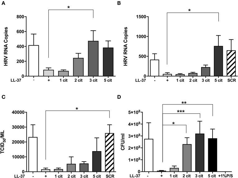
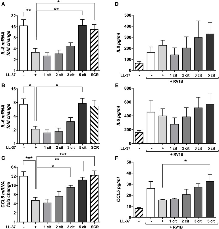
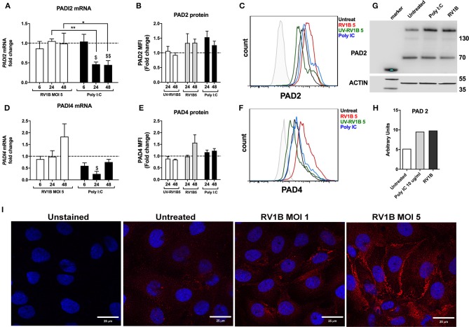
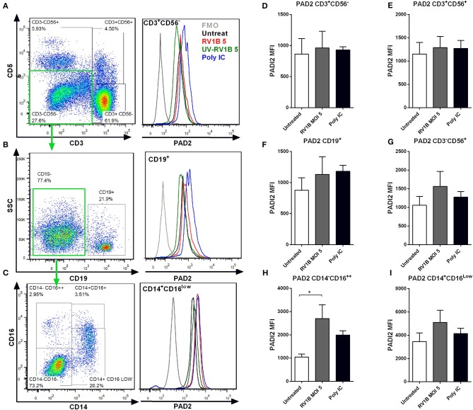
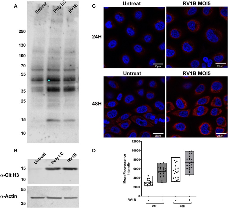
Human rhinoviruses (HRV) are the most common cause of viral respiratory tract infections. While normally mild and self-limiting in healthy adults, HRV infections are associated with bronchiolitis in infants, pneumonia in immunocompromised patients, and exacerbations of asthma and COPD. The human cathelicidin LL-37 is a host defense peptide (HDP) with broad immunomodulatory and antimicrobial activities that has direct antiviral effects against HRV. However, LL-37 is known to be susceptible to the enzymatic activity of peptidyl arginine deiminases (PAD), and exposure of the peptide to these enzymes results in the conversion of positively charged arginines to neutral citrullines (citrullination). Here, we demonstrate that citrullination of LL-37 reduced its direct antiviral activity against HRV. Furthermore, while the anti-rhinovirus activity of LL-37 results in dampened epithelial cell inflammatory responses, citrullination of the peptide, and a loss in antiviral activity, ameliorates this effect. This study also demonstrates that HRV infection upregulates PAD2 protein expression, and increases levels of protein citrullination, including histone H3, in human bronchial epithelial cells. Increased PADI gene expression and HDP citrullination during infection may represent a novel viral evasion mechanism, likely applicable to a wide range of pathogens, and should therefore be considered in the design of therapeutic peptide derivatives.

Keywords: LL-37; cathelicidin; citrullination; inflammation; peptide; rhinovirus; virus.


Figures

Figure 1
Figure 1
Citrullination of LL-37 abrogates the antiviral activity of the peptide toward human rhinovirus. Purified HRV1B viral particles were pre-treated with 10 μg (A) or 30 μg (B) of native or differentially citrullinated LL-37 peptides (Table 1) for 2 h before incubation with 16HBE14°− cells for 1 h at a final MOI of 1. Cells were washed to remove inoculum and incubated for 24 h before viral RNA in cells was addressed by qPCR. To measure released viral particles, cell supernatants from (B) were serially diluted and exposed to WI-38 cells to obtain an infectious viral titer (TCID50/ml) (C). S. aureus bacteria was treated directly with 30 μg/ml of the native and modified LL-37 peptides for 2 h and incubated for 24 h before enumeration of colony formation units (CFU/ml) (D). Data represent mean values ± SEM for n = 3 experiments for each condition *p ≤ 0.05, **p ≤ 0.01, ***p ≤ 0.001. Statistical analysis performed by one-way ANOVA with Dunnet multiple comparisons test.
Figure 2
Figure 2
Citrullination of LL-37 modulates cytokine expression and secretion induced by human rhinovirus. Purified HRV1B viral particles were pre-treated with 30 μg (A–C) or 10 μg (D–F) of native or differentially citrullinated LL-37 peptides for 2 h and incubated with 16HBE14°− cells for 1 h at a final MOI of 1. Cells were washed to remove inoculum and left 24 h before RNA extraction. Levels of IL-8 (A), IL-6 (B), and CCL5 (C) mRNA transcripts were addressed by qPCR. Values represent fold-changes over uninfected cells. Protein concentrations of IL-8 (D), IL-6 (E), and CCL5 (F) released into cell supernatants were assessed by ELISA. Data represent mean values ± SEM for n = 3 experiments for each condition. *p ≤ 0.05, **p ≤ 0.01, ***p ≤ 0.001. Statistical analysis performed by one-way ANOVA with Dunnet multiple comparisons test.
Figure 3
Figure 3
Human rhinovirus and Poly I:C stimulation increase PAD2 protein expression in lung epithelial cells. Human bronchial epithelial (16HBE14°−) cells were infected with HRV1B MOI = 5 for 6, 24 and 48 h before mRNA levels of PADI2 (A) and PADI4 (D) were assessed by qPCR. Poly I:C was used as positive control for viral dsRNA. Protein levels of PAD2 (B) and PAD4 (E) at 24 or 48 h after RV1B infection were measured by FACS (PAD2 and PAD 4) and Western immunoblotting (PAD 2 only, 24 h post infection). UV irradiated virus (UV-RV1B) was used as replication deficient virus control. Values represent fold change expression over untreated cells (dotted line) with data representing the mean ± SEM of 4 different experiments. Representative histogram plots for PAD2 (C) or PAD4 (F) protein expression in 16HBE14°− are shown. A representative Western immunoblot (representative of n = 3) together with quantification by densitometry are displayed (G,H), showing enhanced PAD2 protein in 16HBE14°− cells after 24 h of RV1B infection at MOI = 5. Confocal microscopy images show enhanced PAD2 staining (red) in 16HBE14°− cells after 48 h of RV1B infection at MOI = 1 or MOI = 5 (I). Statistical analysis in (A,D) was performed on ΔΔCT values via a two-way ANOVA with Tukey multiple comparisons test. $p ≤ 0.05, $$p ≤ 0.01 denoting significance on Poly I:C treatment vs. untreated (24 and 48 h, respectively). *p ≤ 0.05, **p ≤ 0.01.
Figure 4
Figure 4
Human rhinovirus and Poly I:C stimulation increase PAD2 expression in CD14CD16++ PBMCs. Peripheral blood mononuclear cells (PBMC) were isolated and exposed to different doses of HRV (viral MOI of 1 or 5) or to 10 μg/ml of Poly I:C for 24 or 48 h. Intracellular PAD2 expression was assessed in different PBMC subsets by FACS. Dot plots indicate the gating strategy used and histogram overlays indicate representative PAD2 levels (MFI) in different PBMCs subsets, (A) CD3+ CD56 T-cells, (B) CD19+ CD3 CD56 B-cells, and (C) CD14+ CD16low CD19 CD3 CD56 Monocytes. Bars represent the Mean Fluorescence Index (MFI) of PAD2 levels expressed in each PBMC subset after 48 h of infection: (D) T-cells, (E) NK-T cells, (F) NK-cells, (G) B-cells, (H) CD14 CD16++ monocytes, and (I) CD14+ CD16low monocytes. Bars indicate the mean ± SEM of three different experiments. Statistical analysis was performed by a one way ANOVA with Tukey's multiple comparisons test (*p ≤ 0.05).
Figure 5
Figure 5
Human rhinovirus and Poly I:C stimulation increase protein citrullination in 16HBE14°− cells. Human bronchial epithelial (16HBE14°−) cells were infected with HRV1B MOI = 5 or treated with Poly IC and total citrullination was measured with a monoclonal antibody against peptidyl citrulline (F95). (A) After 24 h, lysates were obtained and blotted with F95 antibody, α-citrullinated histone H3, and actin, which was used as loading control. (B) Confocal microscopy images were also taken at 24 or 48 h after infection, showing F95 staining (RED) and DAPI as nuclear counterstaining (BLUE). (C) Quantification of at least four different fields of view from (C) is shown in (D), with each dot representing a different cell and boxes displaying min to max and mean values of F95 staining intensity.

References

    1. Jartti T, Gern JE. Rhinovirus-associated wheeze during infancy and asthma development. Curr Respir Med Rev. (2011) 7:160–6. 10.2174/157339811795589423 - DOI - PMC - PubMed
    1. Jacobs SE, Lamson DM, St., George K, Walsh TJ. Human rhinoviruses. Clin Microbiol Rev. (2013) 26:135–62. 10.1128/CMR.00077-12 - DOI - PMC - PubMed
    1. Ruuskanen O, Lahti E, Jennings LC, Murdoch DR. Viral pneumonia. Lancet. (2011) 377:1264–75. 10.1016/S0140-6736(10)61459-6 - DOI - PMC - PubMed
    1. Johnston SL, Pattemore PK, Sanderson G, Smith S, Lampe F, Josephs L, et al. . Community study of role of viral infections in exacerbations of asthma in 9-11 year old children. BMJ. (1995) 310:1225–9. 10.1136/bmj.310.6989.1225 - DOI - PMC - PubMed
    1. Jackson DJ, Gangnon RE, Evans MD, Roberg KA, Anderson EL, Pappas TE, et al. . Wheezing rhinovirus illnesses in early life predict asthma development in high-risk children. Am J Respir Crit Care Med. (2008) 178:667–72. 10.1164/rccm.200802-309OC - DOI - PMC - PubMed

Publication types

LinkOut - more resources