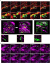
Intravital Microscopy of the Beating Murine Heart to Understand Cardiac Leukocyte Dynamics
- PMID: 32117249
- PMCID: PMC7010807
- DOI: 10.3389/fimmu.2020.00092
Intravital Microscopy of the Beating Murine Heart to Understand Cardiac Leukocyte Dynamics
Abstract
Cardiovascular disease is the leading cause of worldwide mortality. Intravital microscopy has provided unprecedented insight into leukocyte biology by enabling the visualization of dynamic responses within living organ systems at the cell-scale. The heart presents a uniquely dynamic microenvironment driven by periodic, synchronous electrical conduction leading to rhythmic contractions of cardiomyocytes, and phasic coronary blood flow. In addition to functions shared throughout the body, immune cells have specific functions in the heart including tissue-resident macrophage-facilitated electrical conduction and rapid monocyte infiltration upon injury. Leukocyte responses to cardiac pathologies, including myocardial infarction and heart failure, have been well-studied using standard techniques, however, certain questions related to spatiotemporal relationships remain unanswered. Intravital imaging techniques could greatly benefit our understanding of the complexities of in vivo leukocyte behavior within cardiac tissue, but these techniques have been challenging to apply. Different approaches have been developed including high frame rate imaging of the beating heart, explantation models, micro-endoscopy, and mechanical stabilization coupled with various acquisition schemes to overcome challenges specific to the heart. The field of cardiac science has only begun to benefit from intravital microscopy techniques. The current focused review presents an overview of leukocyte responses in the heart, recent developments in intravital microscopy for the murine heart, and a discussion of future developments and applications for cardiovascular immunology.
Keywords: cardiovascular; heart; intravital microscopy; leukocyte; multiphoton microscopy.
Copyright © 2020 Allan-Rahill, Lamont, Chilian, Nishimura and Small.
Figures


Similar articles
-
Intravital Imaging of the Heart at the Cellular Level Using Two-Photon Microscopy.Methods Mol Biol. 2018;1763:145-151. doi: 10.1007/978-1-4939-7762-8_14. Methods Mol Biol. 2018. PMID: 29476496
-
Frontiers in Intravital Multiphoton Microscopy of Cancer.Cancer Rep (Hoboken). 2020 Feb;3(1):e1192. doi: 10.1002/cnr2.1192. Epub 2019 Jun 20. Cancer Rep (Hoboken). 2020. PMID: 32368722 Free PMC article. Review.
-
Visualizing murine breast and melanoma tumor microenvironment using intravital multiphoton microscopy.STAR Protoc. 2021 Aug 19;2(3):100722. doi: 10.1016/j.xpro.2021.100722. eCollection 2021 Sep 17. STAR Protoc. 2021. PMID: 34458865 Free PMC article.
-
Intravital Imaging of Liver Cell Dynamics.Methods Mol Biol. 2018;1763:137-143. doi: 10.1007/978-1-4939-7762-8_13. Methods Mol Biol. 2018. PMID: 29476495
-
Intravital 2-photon imaging, leukocyte trafficking, and the beating heart.Trends Cardiovasc Med. 2013 Nov;23(8):287-93. doi: 10.1016/j.tcm.2013.04.002. Epub 2013 May 22. Trends Cardiovasc Med. 2013. PMID: 23706535 Free PMC article. Review.
Cited by
-
Viscocohesive hyaluronan gel enhances stability of intravital multiphoton imaging with subcellular resolution.Neurophotonics. 2025 Jan;12(Suppl 1):S14602. doi: 10.1117/1.NPh.12.S1.S14602. Epub 2024 Nov 22. Neurophotonics. 2025. PMID: 39583344 Free PMC article.
-
Editorial: Community series in intravital microscopy imaging of leukocytes, volume II.Front Immunol. 2025 May 9;16:1615392. doi: 10.3389/fimmu.2025.1615392. eCollection 2025. Front Immunol. 2025. PMID: 40416958 Free PMC article. No abstract available.
-
Intravital Imaging Allows Organ-Specific Insights Into Immune Functions.Front Cell Dev Biol. 2021 Feb 11;9:623906. doi: 10.3389/fcell.2021.623906. eCollection 2021. Front Cell Dev Biol. 2021. PMID: 33644061 Free PMC article. Review.
-
Intravital imaging of the functions of immune cells in the tumor microenvironment during immunotherapy.Front Immunol. 2023 Dec 6;14:1288273. doi: 10.3389/fimmu.2023.1288273. eCollection 2023. Front Immunol. 2023. PMID: 38124754 Free PMC article. Review.
-
Genetically engineered mice for combinatorial cardiovascular optobiology.Elife. 2021 Oct 29;10:e67858. doi: 10.7554/eLife.67858. Elife. 2021. PMID: 34711305 Free PMC article.
References
-
- Klabunde RE. Myocardial oxygen demand. In: Taylor C. editor. Cardiovascular Physiology Concepts. 2nd ed Baltimore, MD: Lippincott Williams & Wilkins; (2011), p. 182–6.
-
- GBD 2017 Causes of Death Collaborators. Global, regional, and national age-sex-specific mortality for 282 causes of death in 195 countries and territories, 1980-2017: a systematic analysis for the Global Burden of Disease Study 2017. Lancet. (2018) 392:1736–88. 10.1016/S0140-6736(18)32203-7 - DOI - PMC - PubMed
Publication types
MeSH terms
Grants and funding
LinkOut - more resources
Full Text Sources

