. 2020 Feb 14:11:108.
doi: 10.3389/fimmu.2020.00108. eCollection 2020.

IL-17A Modulates Peritoneal Macrophage Recruitment and M2 Polarization in Endometriosis

Affiliations

IL-17A Modulates Peritoneal Macrophage Recruitment and M2 Polarization in Endometriosis

Jessica E Miller et al. Front Immunol. .

Abstract

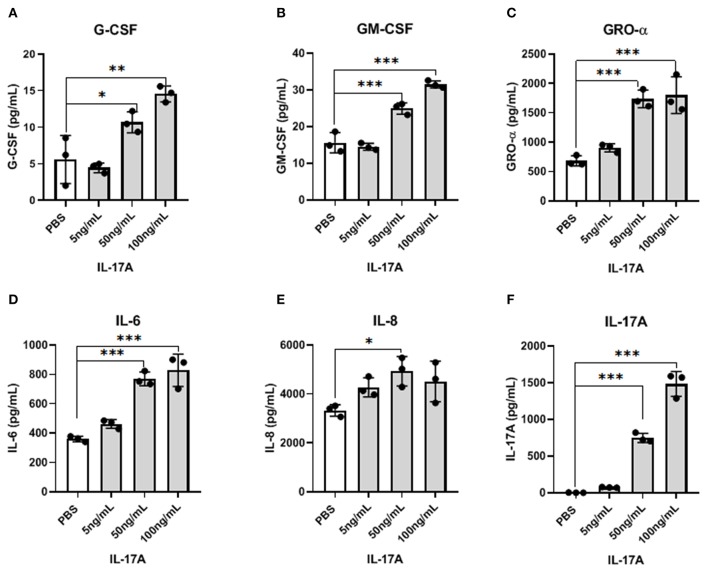
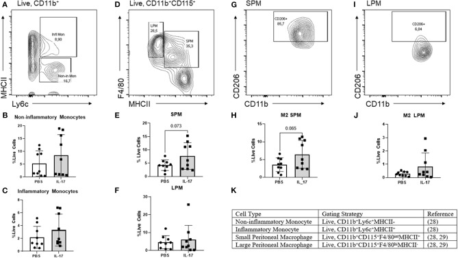
Endometriosis is a debilitating gynecological disease characterized by the extrauterine presence of endometrial-like tissues located on the peritoneal membrane and organs of the pelvic cavity. Notably, dysfunctional immune activation in women with endometriosis could also contribute to the development of disease. In particular, alternatively activated (M2) peritoneal macrophages are shown to aid peritoneal lesion development by promoting remodeling of extracellular matrix and neovascularization of lesions. However, the stimuli responsible for polarizing M2 macrophages in endometriosis remain elusive. Interleukin-17A (IL-17A) can induce M2 macrophage polarization in other disease models and IL-17A is elevated in the plasma and endometriotic lesions of women with endometriosis. In this study, we investigated whether IL-17A could induce macrophage recruitment and M2 polarization, while promoting endometriotic lesion growth through enhanced vascularization. By utilizing a co-culture of macrophage-like THP-1 cells with an endometriotic epithelial cell line, our in vitro results suggest that IL-17A indirectly induces M2 markers CCL17 and CD206 by interacting with endometriotic epithelial cells. Further, in a syngeneic mouse model of endometriosis, IL-17A treatment increased macrophages in the peritoneum, which were also M2 in phenotype. However, IL-17A treatment did not augment proliferation or vascularization of the lesion in the study time frame. These findings suggest that IL-17A may be a stimulus inducing the pathogenic polarization of macrophages into the M2 phenotype by first acting on the endometriotic lesion itself.

Keywords: M2 macrophage; cytokines; endometriosis; inflammation; interleukin-17A.


Figures

Figure 1
Figure 1
IL-17A induces IL1B expression and, GRO- α and G-CSF production in PMA-differentiated THP-1 cells. THP-1 cells were differentiated into macrophages using PMA (10 ng/mL) for 48 h followed by IL-17A (50 ng/mL) stimulation for 24 h in PMA-free media. Total RNA was collected from differentiated THP-1 cells treated with either without PMA and with PMA and treated with either PBS or IL-17A. Semi-quantitative RT-PCR revealed IL1B to be upregulated in IL-17A-treated cells (A). In the supernatant collected following 24 h incubation with IL-17A, we detected increased concentration of GRO-α (B), G-CSF (C) and IL-17A (D) in differentiated THP-1 cells (gray columns) compared to non-differentiated THP-1 cells (white columns). Statistical analysis conducted using One-way ANOVA with Bonferroni post-hoc test. *P ≤ 0.05, **P ≤ 0.01, ***P ≤ 0.001.
Figure 2
Figure 2
IL-17A induces the production of inflammatory cytokines and chemokines from 12Z cells in a dose-dependent manner. 12Z cells were plated in 6 wells at 1 × 10⁶/ well and incubated for 24 h with different concentration of IL-17A (5 ng/mL, 50 ng/mL, and 100 ng/mL), as well as with PBS as control. Out of 42 cytokines analyzed in the supernatant, G-CSF (A), GM-CSF (B), GRO-α (C), IL-6 (D), and IL-8 (E) showed dose-dependent increase in concentration that was significantly different from the level obtained with PBS treatment. Statistical analysis conducted using one-way ANOVA with Tukey post-hoc test (A–F). *P ≤ 0.05, **P ≤ 0.01, ***P ≤ 0.001.
Figure 3
Figure 3
THP-1 cells incubated with conditioned media from 12Z cells treated with IL-17A showed alterations in M2 polarization markers. 12Z cells were plated at 1 x 10⁶ cells per well and incubated for 24 h with either PBS or 50 ng/mL of IL-17A. THP-1 cells were then incubated with this conditioned media for 24 h. Total RNA was extracted from THP-1 cells to test for M1 and M2 markers via RT-qPCR. Marker for M2 macrophages, ccl17 (A) was significantly upregulated in THP-1 cells treated with IL-17A-CM compared to PBS-CM. Transcription level of CD206 (B), also a marker of M2 macrophage, was increased in IL-17A-CM treated cells; however, the difference did not reach statistical significance. Statistical analysis conducted using an unpaired t-test with Welch's correction (A,B). *P ≤ 0.05.
Figure 4
Figure 4
Bone marrow derived macrophages treated with IL-17 secreted elevated levels of macrophage associated cytokines however showed no differences in M2 polarization status compared to PBS-treated controls. (A) Histogram showing no difference in CD206 expression between BMDMs (Live, CD11b+, F4/80+, CD115+ cells) treated with PBS compared to treated with 5, 50, and 100 ng/mL of rmIL-17. (B) No significant differences were found in CD206+ BMDMs treated with IL-17 compared to PBS controls. Out of 31 analytes, IL-17 (C), GRO-α (D), MIP-1α (E), and MIP-1β (F) were found to be significantly elevated in the supernatant of IL-17 treated BMDMs compared to PBS controls. Statistical analysis conducted using one-way ANOVA with Tukey post-hoc test (B–F). *P ≤ 0.05, **P ≤ 0.01, ***P ≤ 0.001.
Figure 5
Figure 5
Characterization of monocytes and macrophages in the peritoneal lavage fluid of mice treated with IL-17A or PBS. (A,D,G,I,K) Gating strategy for monocytes, non-inflammatory monocytes, inflammatory monocytes, small peritoneal macrophage (SPM), large peritoneal macrophage (LPM) and CD206 polarization status. (B,C) Scatter plot for non-inflammatory and inflammatory monocytes shows no difference between IL-17A (n = 9) and PBS group (n = 9). (E) Scatter plot for % Live cells peritoneal lavage fluid shows elevated SPM in the IL-17A (n = 9) treated group compared to PBS group (n = 9). (F) No difference was found in the populations of LPM. (H,J) SPM had elevated CD206 in the IL-17A-treated group (n = 9) compared to the PBS-treated group (n = 9). No significant differences in CD206 were found in LPM. Scatter plot for % F4/80+CD206+ cells from CD11bhi population show no difference between IL-17A (n = 9) and PBS group (n = 9) based on unpaired t-test with Welch's correction.
Figure 6
Figure 6
Multiplex cytokine analysis in plasma samples of mice treated with IL-17A (0.5 μg/100 μl). Out of 31 analytes assessed, Eotaxin (A), IL-9 (B), IL-13 (C), and MIP-α (D) were decreased in plasma after 1 week of both PBS (n = 4) and IL-17A (n = 4) treatment. Repeated measurement 2-way ANOVA with Sidak multiple comparison correction method was used as statistical analysis. *P ≤ 0.05, **P ≤ 0.01.
Figure 7
Figure 7
Multiplex cytokine analysis in peritoneal fluid of mice treated with IL-17A (0.5 μg/100 μl). Out of 31 analytes assessed, IL-9 (A), IL-10 (B), IL-15 (C), IL-17A (D), CCL11/Eotaxin (E), CXCL1/KC (F), and G-CSF (G) were detected; however, no significant differences were found between PBS (n = 4) and IL-17 (n = 4) treated mice. Undetected analytes are not shown. Unpaired t-test was used as statistical analysis. *P ≤ 0.05.
Figure 8
Figure 8
Gross and histological images of endometriosis lesions in mice treated with IL-17A or PBS. PBS (A; n = 4) and IL-17A (B, n = 4). Both PBS-treated (C) and IL-17A-treated (D) lesions had Ki67+ cells within the stromal region of the tissue, with occasional positivity in the luminal epithelium. Semi quantitative analysis revealed no significant differences in Ki67 staining. Both IL-17A-treated (F) and PBS control lesions (G) show extensive vascularization within the stroma as denoted by cross sections of vessels stained in brown. Semi-quantitative analysis showed no significant difference (E,H). All images are taken at 200x magnification.
Figure 9
Figure 9
Visual Summary: IL-17A is involved in peritoneal M2 polarization in endometriosis.

References

    1. Giudice LC. Clinical practice. Endometriosis. N Engl J Med. (2010) 362:2389–98. 10.1056/NEJMcp1000274 - DOI - PMC - PubMed
    1. D'Hooghe TM, Bambra CS, Suleman MA, Dunselman GA, Evers HL, Koninckx PR. Development of a model of retrograde menstruation in baboons (Papio anubis)**Supported by the Commission of the European Communities (DG VIII Development and DG XII Science, Research and Development) and by the Vlaamse Interuniversitaire Raad (Flemish Interuniversity Council), Brussels, Belgium. Fertil Steril. (1994) 62:635–8. 10.1016/S0015-0282(16)56957-X - DOI - PubMed
    1. Sampson JA. Peritoneal endometriosis due to the menstrual dissemination of endometrial tissue into the peritoneal cavity. Am J Obstet Gynecol. (1927) 14:422–69. 10.1016/S0002-9378(15)30003-X - DOI - PubMed
    1. Capobianco A, Rovere-Querini P. Endometriosis, a disease of the macrophage. Front Immunol. (2013) 4:9. 10.3389/fimmu.2013.00009 - DOI - PMC - PubMed
    1. McLaren J, Prentice A, Charnock-Jones DS, Millican SA, Müller KH, Sharkey AM, et al. . Vascular endothelial growth factor is produced by peritoneal fluid macrophages in endometriosis and is regulated by ovarian steroids. J Clin Invest. (1996) 98:482–9. 10.1172/JCI118815 - DOI - PMC - PubMed

Publication types

MeSH terms

Grants and funding

LinkOut - more resources