. 2020 Feb 13:11:155.
doi: 10.3389/fimmu.2020.00155. eCollection 2020.

Microbial Regulation of Enteric Eosinophils and Its Impact on Tissue Remodeling and Th2 Immunity

Affiliations

Microbial Regulation of Enteric Eosinophils and Its Impact on Tissue Remodeling and Th2 Immunity

Rodrigo Jiménez-Saiz et al. Front Immunol. .

Abstract

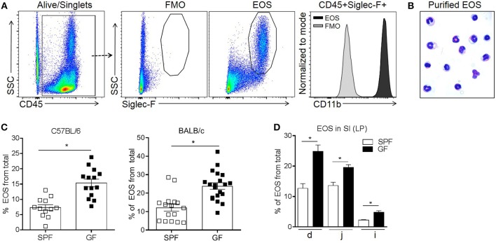
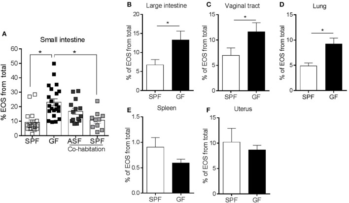
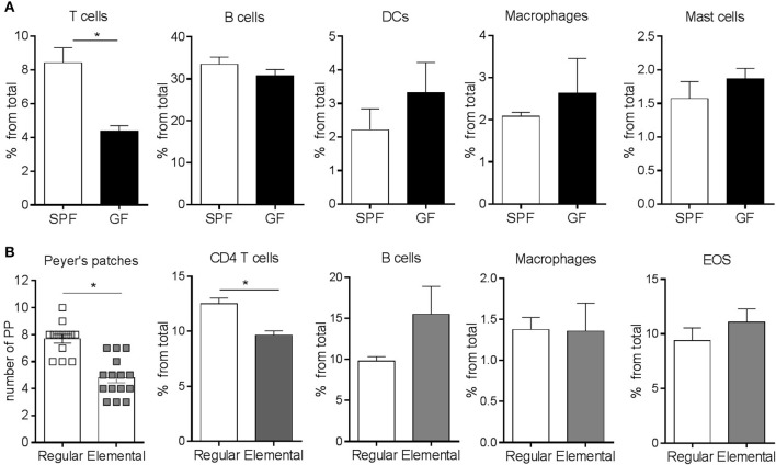
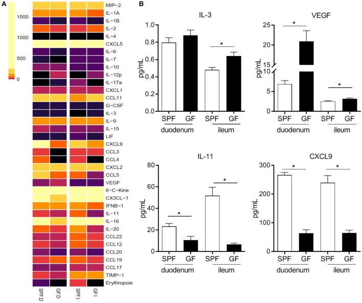
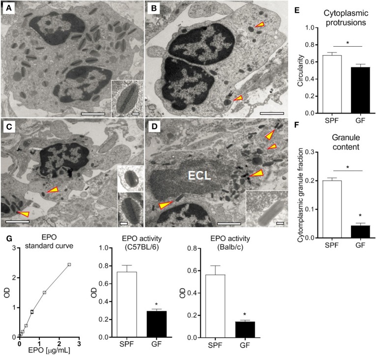
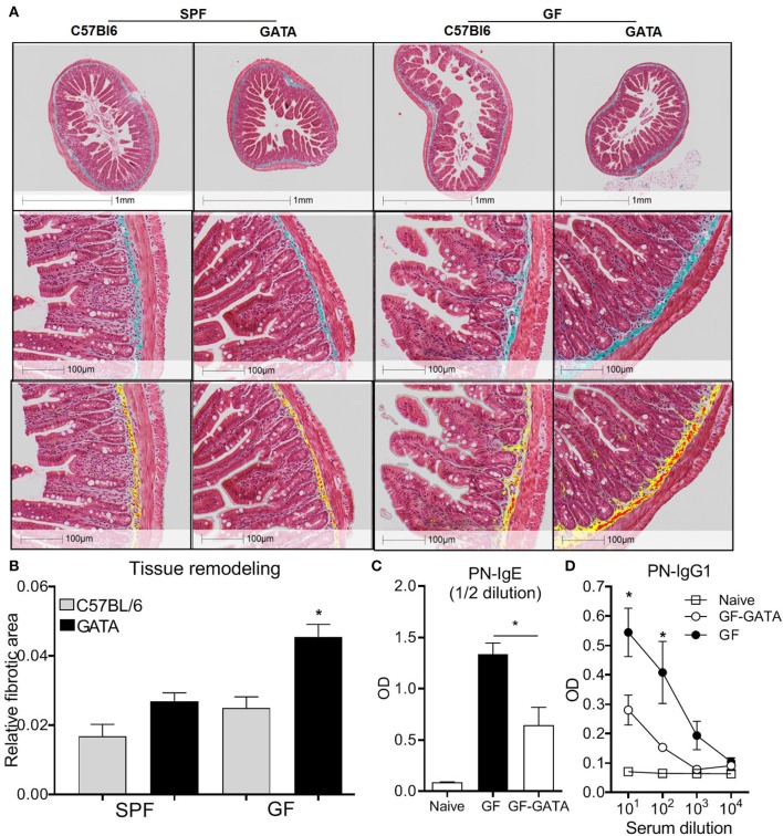
Eosinophils have emerged as multifaceted cells that contribute to tissue homeostasis. However, the impact of the microbiota on their frequency and function at mucosal sites remains unclear. Here, we investigated the role of the microbiota in the regulation of enteric eosinophils. We found that small intestinal (SI) eosinophilia was significantly greater in germ-free (GF) mice compared to specific pathogen free (SPF) controls. This was associated with changes in the production of enteric signals that regulate eosinophil attraction and survival, and was fully reversed by complex colonization. Additionally, SI eosinophils of GF mice exhibited more cytoplasmic protrusions and less granule content than SPF controls. Lastly, we generated a novel strain of eosinophil-deficient GF mice. These mice displayed intestinal fibrosis and were less prone to allergic sensitization as compared to GF controls. Overall, our study demonstrates that commensal microbes regulate intestinal eosinophil frequency and function, which impacts tissue repair and allergic sensitization to food antigens. These data support a critical interplay between the commensal microbiota and intestinal eosinophils in shaping homeostatic, innate, and adaptive immune processes in health and disease.

Keywords: IgE; allergy; eosinophils; germfree; microbiota; small intestine; tissue remodeling.


Figures

Figure 1
Figure 1
Flow cytometric identification of small intestinal eosinophils (EOS) as live singlet CD45+Siglec-F+ cells (A), morphologic validation (B) and assessment of their frequency in the small intestine (SI) of SPF and GF C57BL/6 and BALB/c mice (C,D). The frequency of EOS from total cells in the lamina propria (LP) of different sections of the SI including duodenum (d), jejunum (j) and ileum (i) of C57BL/6 mice (D). Pooled data from 3 to 4 experiments (n = 12–20) (C) or representative data from 3 experiments (D) represented as mean ± SEM, *P < 0.05.
Figure 2
Figure 2
Separate groups of GF mice were colonized by co-habitation with ASF or SPF mice and the presence of small intestinal eosinophils (EOS) was assessed (A). Assessment of EOS frequency in the large intestine, vaginal tract, lung, spleen and uterus of SPF and GF mice by flow cytometry (B–F). Pooled data from 2 to 5 experiments (n = 8–22) represented as mean ± SEM, *P < 0.05.
Figure 3
Figure 3
Flow cytometric characterization of adaptive and innate immune compartments (A,B) in the small intestine of GF and SPF mice fed a regular (A) or elemental diet (B). Pooled data from 2 to 4 experiments (n = 6–22) represented as mean ± SEM, *P < 0.05.
Figure 4
Figure 4
Heatmap of chemokine and cytokine protein levels in proximal (duodenum; D) and distal (ileum; I) small intestinal homogenates from GF and SPF mice as determined by protein array (A). Relevant proteins differentially produced both in the proximal and distal small intestine of SPF and GF mice (B). Data from 6 mice (A,B) represented as mean ± SEM, *P < 0.05.
Figure 5
Figure 5
Normal SI eosinophils transmission electron microscopy ultrastructure (A), showing bi-lobed nuclei and a high density of granules composed of an electron-dense core surrounded by an electron-lucent matrix rich in EPO (arrowhead). SI eosinophils from GF mice (B–D) exhibit cytoplasmic protrusions, measured as reduced circularity (E), lower content of cytoplasmic granules (F) and, occasionally, ECL signs (D). Assessment of EPO in intestinal homogenates of SPF and GF mice (G). Representative (A–D) and pooled (E,F) data from 6 mice, and pooled data from 2 independent experiments (n = 6) (G) represented as mean ± SEM, *P < 0.05.
Figure 6
Figure 6
Representative histological examples of intestinal fibrosis (A, upper panels) and associated Halo quantification of fibrotic surface on digitalized (20X) sections where yellow and red represents Masson's Trichrome positive stain (A, lower panels) and associated quantification (B) of overall fibrotic area performed on GF mice, eosinophil deficient GF mice (GATA-GF) and controls. GF mice, eosinophil deficient GF mice (GATA-GF) and controls were intragastrically sensitized to peanut (PN) and serum levels of PN-specific IgE (C) and IgG1 (D) were determined by ELISA. Data from 4 to 5 mice (A,B) or pooled data from 2 experiments (n = 6) (C,D) represented as mean ± SEM, *P < 0.05.

References

    1. Lee JJ, Jacobsen EA, McGarry MP, Schleimer RP, Lee NA. Eosinophils in health and disease: the LIAR hypothesis. Clin Exp Allergy. (2010) 40:563–75. 10.1111/j.1365-2222.2010.03484.x - DOI - PMC - PubMed
    1. Rosenberg HF, Dyer KD, Foster PS. Eosinophils: changing perspectives in health and disease. Nat Rev Immunol. (2013) 13:9–22. 10.1038/nri3341 - DOI - PMC - PubMed
    1. Arnold IC, Artola-Boran M, Tallon de Lara P, Kyburz A, Taube C, Ottemann K, et al. . Eosinophils suppress Th1 responses and restrict bacterially induced gastrointestinal inflammation. J Exp Med. (2018) 215:2055–72. 10.1084/jem.20172049 - DOI - PMC - PubMed
    1. Abdala-Valencia H, Coden ME, Chiarella SE, Jacobsen EA, Bochner BS, Lee JJ, et al. . Shaping eosinophil identity in the tissue contexts of development, homeostasis, and disease. J Leukoc Biol. (2018) 104:95–108. 10.1002/JLB.1MR1117-442RR - DOI - PMC - PubMed
    1. Xenakis JJ, Howard ED, Smith KM, Olbrich CL, Huang Y, Anketell D, et al. . Resident intestinal eosinophils constitutively express antigen presentation markers and include two phenotypically distinct subsets of eosinophils. Immunology. (2018) 154:298–308. 10.1111/imm.12885 - DOI - PMC - PubMed

Publication types

MeSH terms

Grants and funding