. 2020 Apr 1;9(4):145-160.
doi: 10.1089/wound.2019.0956. Epub 2020 Feb 7.

Circular RNA hsa_circ_0084443 Is Upregulated in Diabetic Foot Ulcer and Modulates Keratinocyte Migration and Proliferation

Affiliations

Circular RNA hsa_circ_0084443 Is Upregulated in Diabetic Foot Ulcer and Modulates Keratinocyte Migration and Proliferation

Aoxue Wang et al. Adv Wound Care (New Rochelle). .

Abstract

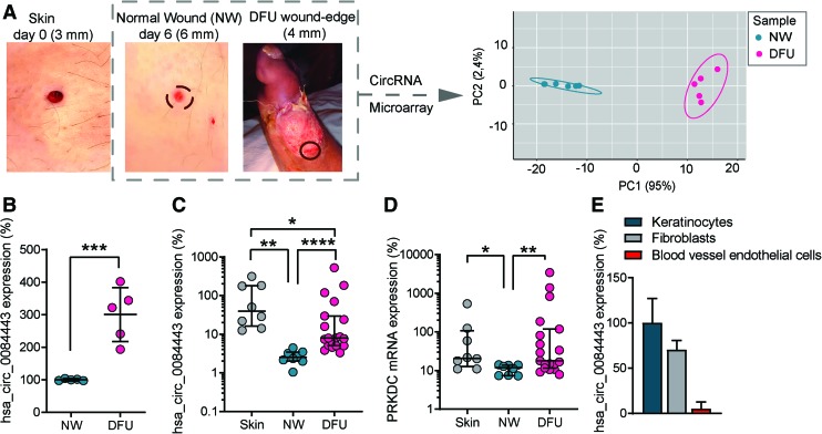
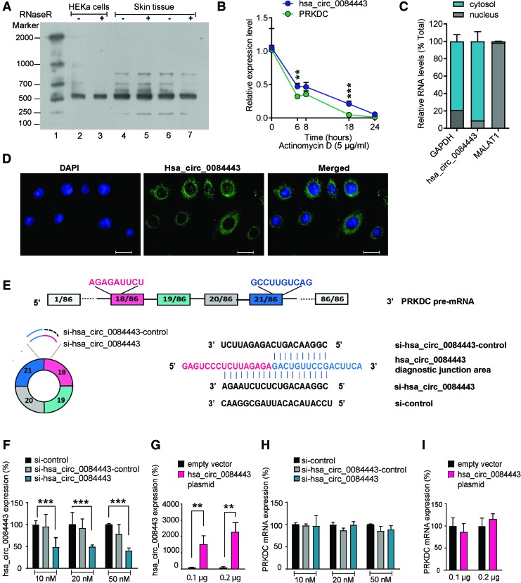
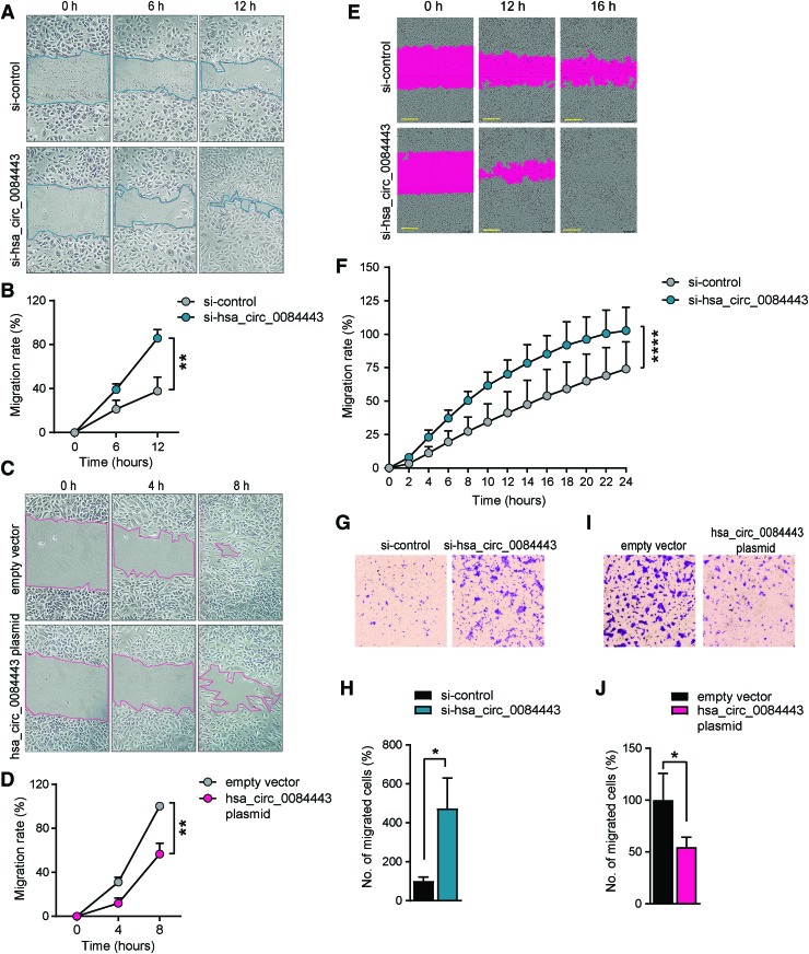
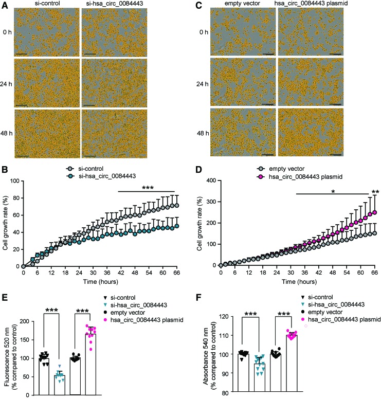
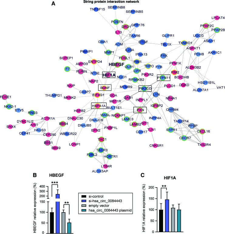
Objective: Insufficient knowledge about the molecular pathology of diabetic foot ulcer (DFU) impedes the development of effective wound treatment. Circular RNAs (circRNAs) are a novel class of RNA recently discovered to be widely expressed and have important biological functions; however, their role in skin wound healing remains largely unexplored. In this study, we investigated the role of circRNAs in DFU. Approach: CircRNA expression was profiled in normal wounds (NWs) and DFUs by microarray analysis, and hsa_circ_0084443 was identified as differentially expressed. The circularity and subcellular localization of hsa_circ_0084443 were characterized by northern blotting, real-time PCR, and fluorescence in situ hybridization. Cell migration, cell growth, and the transcriptome of human primary keratinocytes were analyzed after overexpression or RNA interference of hsa_circ_0084443. Results: hsa_circ_0084443 is downregulated in NWs compared with intact skin, and its level is higher in DFUs than NWs. We confirmed its circularity and presence in the cytoplasm of human epidermal keratinocytes. We showed that hsa_circ_0084443 reduced motility while enhancing the growth of keratinocytes. Furthermore, we identified a gene network with the potential to mediate the biological effect of hsa_circ_0084443. Innovation: CircRNAs have a functional role and a potential clinical significance in skin wound healing. Conclusions: We identified hsa_circ_0084443, a circRNA downregulated during NW healing, as a negative regulator of keratinocyte migration. Higher levels of hsa_circ_0084443 were detected in DFU samples, suggesting that it plays a role in pathology. These findings pave the way to understanding the functional role of circRNAs in human skin wound healing.

Keywords: circular RNA; diabetic foot ulcer; keratinocyte; noncoding RNA; wound healing.


Conflict of interest statement

No competing financial interests exist. The content of this article was expressly written by the authors listed. No ghostwriters were used to write this article.

Figures

None
Aoxue Wang, MD, PhD
None
Ning Xu Landén, MD, PhD
Figure 1.
Figure 1.
Circular RNA hsa_circ_0084443 is upregulated in human chronic diabetic wounds. (A) Biopsies (4 mm in diameter) were collected at wound edges of DFU (n = 5). A 3 mm-surgical wound was created on the skin of healthy volunteers (n = 5) and the wound edge was excised with a 6 mm biopsy punch 6 days later (NW). A principal component analysis was performed on the circRNA microarray data of DFU and NW. (B) The normalized expression levels of hsa_circ_0084443 were extracted from the microarray data. qRT-PCR analysis of hsa_circ_0084443 (C) and PRKDC mRNA (D) in skin (n = 8), NW (n = 8), and DFU (n = 19). (E) qRT-PCR analysis of hsa_circ_0084443 in human primary keratinocytes, fibroblasts and blood vessel endothelial cells. *p < 0.05, **p < 0.01, ***p < 0.001, and ****p < 0.0001 by unpaired two-tailed Student's t-test (B) and Wilcoxon matched pairs signed rank test or Mann–Whitney U test (C, D). Data are presented as mean ± SD. (B), median ± interquartile range (IQR) (C, D), and mean + SD. (E). DFU, diabetic foot ulcer; NW, normal wound; qRT-PCR, quantitative real-time polymerase chain reaction. Color images are available online.
Figure 2.
Figure 2.
hsa_circ_0084443 localizes in the cytoplasm of human epidermal keratinocytes and its aberrant expression does not change PRKDC levels. (A) Northern blotting analysis of hsa_circ_0084443 expression in human primary keratinocytes (HEKa) (lane 2, 3) and skin tissue (lane 4–7) with and without RNase R treatment. RNA molecular weight marker was used (lane 1). (B) qRT-PCR analysis of hsa_circ_0084443 and PRKDC mRNA in HEKa treated with Actinomycin D at the indicated timepoints (n = 3). (C) qRT-PCR analysis of hsa_circ_0084443, MALAT1, and GAPDH in nucleus or cytoplasm of HEKa (n = 3). (D) Fluorescence in situ hybridization to visualize hsa_circ_0084443 in HEKa. Scale bar 20 μm. (E) Illustration of hsa_circ_0084443 and pre-mRNA of PRKDC. Sequence information of si-hsa_circ_0084443, si-control, and si-hsa_circ_0084443-control. qRT-PCR of hsa_circ_0084443 (F) and PRKDC mRNA (H) in keratinocytes transfected with 10, 20, and 50 nM si-control, si-hsa_circ_0084443, or si-hsa_circ_0084443-control for 24 h (n = 3). QRT-PCR of hsa_circ_0084443 (G) and PRKDC mRNA (I) in keratinocytes transfected with hsa_circ_0084443 overexpression plasmid (0.1 or 0.2 μg) for 24 h (n = 3) **p < 0.01 and ***p < 0.001 by unpaired two-tailed Student's t test. Data are presented as mean + SD. Color images are available online.
Figure 3.
Figure 3.
hsa_circ_0084443 inhibits keratinocyte migration. Scratch wound assay of keratinocytes transfected with 20nM si-hsa_circ_0084443 and si-control (A, B) or 0.1 μg hsa_circ_0084443 plasmid and empty vector (C, D) for 24 h (n = 4). Photographs were taken at the indicated timepoints after the scratch (A, C). The healing rates were quantified (B, D). Live-cell imaging using the IncuCyte® system of migrating keratinocytes with hsa_circ_0084443 knockdown (n = 12). Representative photographs of migrating keratinocytes at the indicated timepoints. Scale bar = 300 μm (E). The migration rates were quantified by measuring the area of the scratched region (F). Representative photographs of the transwell migration assay for keratinocytes with hsa_circ_0084443 knockdown (n = 3) (G) or hsa_circ_0084443 overexpression (n = 3) (I). The number of cells passing through the membrane was counted (H, J). *p < 0.05, **p < 0.01, and ****p < 0.0001 by unpaired two-tailed Student's t test (B, D, H, J) and by two-way ANOVA (F). Data are presented as mean + SD. Color images are available online.
Figure 4.
Figure 4.
hsa_circ_0084443 promotes keratinocyte growth. Growth of keratinocytes with hsa_circ_0084443 knockdown (A) or hsa_circ_0084443 overexpression (C) was monitored using the IncuCyte Live-Cell Imaging System (n = 5). Scale bar 300 μm. (B, D) The results were quantified and presented as cell growth rate. (E) Keratinocytes were transfected with si-hsa_circ_0084443 or hsa_circ_0084443 overexpression plasmid for 24 h. The number of viable cells was quantified with CyQUANT cell proliferation assay (n = 12). (F) Colony formation assay of keratinocytes with hsa_circ_0084443 knockdown or overexpression (n = 12). *p < 0.05 and ***p < 0.001 by two-way ANOVA (B, D) or by unpaired two-tailed Student's t-test (E, F). Data are presented as mean + SD. Color images are available online.
Figure 5.
Figure 5.
PI3K, EGFR, and ERK signaling pathways are important for the antimigratory function of hsa_circ_0084443 in keratinocytes. Gene expression profile of keratinocytes with hsa_circ_0084443 knockdown (n = 3) was analyzed by microarray. (A) Gene ontology analysis of the genes regulated by hsa_circ_0084443 (absolute fold change ≥1.5, p < 0.05). Live-cell imaging using the IncuCyte system of migrating keratinocytes with hsa_circ_0084443 knockdown (n = 6) and treated with DMSO or Wortmannin (1 μM) (B), PD153035 (500 nM) (C) or U0126 (10 μM) (D). The migration rates were calculated by measuring the area of the scratched region (B–D). *p < 0.05, ***p < 0.001, and ****p < 0.001 by two-way ANOVA. Data are presented as mean + SD. Color images are available online.
Figure 6.
Figure 6.
A gene network regulated by hsa_circ_0084443 in keratinocytes. (A) Functional protein association network was identified by String APP in Cytoscape software among the genes regulated by hsa_circ_0084443 shown in the microarray analysis. Genes up- or downregulated after transfection of si-hsa_circ_0084443 are in red or blue, respectively. Genes previously reported to have an impact on cell migration and proliferation are highlighted: yellow border = proliferation genes, green border = migration genes, grey border = migration and proliferation genes. Core nodes identified by centrality analysis with the CentiScaPe 2.2 App are marked with black rectangles. qRT-PCR of HBEGF mRNA (B) and HIF1A mRNA (C) in keratinocytes with hsa_circ_0084443 knockdown (n = 10) or overexpression (n = 4). **p < 0.01 and ***p < 0.001 by unpaired two-tailed Student's t-test. Data are presented as mean + SD. Color images are available online.

Similar articles

Cited by

References

    1. World Health Organization. Global Report on Diabetes. Geneva, Switzerland: World Health Organization, 2016;978:88
    1. Guo S, DiPietro L. Factors affecting wound healing. J Dent Res 2010;89:219–229 - PMC - PubMed
    1. Frykberg RG, Zgonis T, Armstrong DG, et al. . Diabetic foot disorders. A clinical practice guideline (2006 revision). J Foot Ankle Surg 2006;45:S1–S66 - PubMed
    1. Baltzis D, Eleftheriadou I, Veves A. Pathogenesis and treatment of impaired wound healing in diabetes mellitus: new insights. Adv Ther 2014;31:817–836 - PubMed
    1. Catrina SB, Zheng X. Disturbed hypoxic responses as a pathogenic mechanism of diabetic foot ulcers. Diabetes Metab Res Rev 2016;32:179–185 - PubMed

Publication types