. 2020 Feb 18:8:e8556.
doi: 10.7717/peerj.8556. eCollection 2020.

Broad similarities in shoulder muscle architecture and organization across two amniotes: implications for reconstructing non-mammalian synapsids

Affiliations

Broad similarities in shoulder muscle architecture and organization across two amniotes: implications for reconstructing non-mammalian synapsids

Philip Fahn-Lai et al. PeerJ. .

Abstract

The evolution of upright limb posture in mammals may have enabled modifications of the forelimb for diverse locomotor ecologies. A rich fossil record of non-mammalian synapsids holds the key to unraveling the transition from "sprawling" to "erect" limb function in the precursors to mammals, but a detailed understanding of muscle functional anatomy is a necessary prerequisite to reconstructing postural evolution in fossils. Here we characterize the gross morphology and internal architecture of muscles crossing the shoulder joint in two morphologically-conservative extant amniotes that form a phylogenetic and morpho-functional bracket for non-mammalian synapsids: the Argentine black and white tegu Salvator merianae and the Virginia opossum Didelphis virginiana. By combining traditional physical dissection of cadavers with nondestructive three-dimensional digital dissection, we find striking similarities in muscle organization and architectural parameters. Despite the wide phylogenetic gap between our study species, distal muscle attachments are notably similar, while differences in proximal muscle attachments are driven by modifications to the skeletal anatomy of the pectoral girdle that are well-documented in transitional synapsid fossils. Further, correlates for force production, physiological cross-sectional area (PCSA), muscle gearing (pennation), and working range (fascicle length) are statistically indistinguishable for an unexpected number of muscles. Functional tradeoffs between force production and working range reveal muscle specializations that may facilitate increased girdle mobility, weight support, and active stabilization of the shoulder in the opossum-a possible signal of postural transformation. Together, these results create a foundation for reconstructing the musculoskeletal anatomy of the non-mammalian synapsid pectoral girdle with greater confidence, as we demonstrate by inferring shoulder muscle PCSAs in the fossil non-mammalian cynodont Massetognathus pascuali.

Keywords: Forelimb; Locomotion; Mammals; Musculoskeletal function; Non-mammalian synapsid; Posture; Shoulder.


Conflict of interest statement

Stephanie E. Pierce is an Academic Editor for PeerJ.

Figures

Figure 1
Figure 1. Skeletal anatomy and midstance posture of (A and B) “sprawling” Salvator merianae, (C and D) Massetognathus pascuali, and (E and F) “erect” Didelphis virginiana pectoral limb.
All animals shown in right lateral view (A, C, E) and cranial view (B, D, F). M. pascuali is depicted in a hypothetical neutral pose, reflecting the middle of each joint’s range of motion (see Lai, Biewener & Pierce, 2018). S. merianae and D. virginiana poses taken from 3D videoradiography. While in Massetognathus the ventral portion of the scapulocoracoid comprises separate procoracoid and metacoracoid elements (sensu Vickaryous & Hall, 2006), here they are collectively termed the “coracoid” for simplicity.
Figure 2
Figure 2. Right lateral view of pectoral girdle and proximal forelimb of the Argentine black and white tegu (Salvator merianae) (A and B) and the Virginia opossum (Didelphis virginiana) (C and D).
Showing all muscles crossing the shoulder joint (A and C), and the deep layer of muscles originating on the scapulocoracoid/scapula (B and D).
Figure 3
Figure 3. Muscle attachments on the right scapulocoracoid/scapula of Salvator merianae (s) and Didelphis virginiana (d).
Shown in medial (A and B), caudal (C and D), cranial (E and F), and lateral (G and H) view. Stippled areas represent loose fascial associations between muscle and bone. Muscle abbreviations and color-coding follow Fig. 2.
Figure 4
Figure 4. Muscle attachments on the right humerus of Salvator merianae (s) and Didelphis virginiana (d).
Shown in medial (A and B), extensor (C and D), flexor (E and F), and lateral (G and H) view. Muscle abbreviations and color-coding follow Fig. 2.
Figure 5
Figure 5. Muscle attachments on the right clavicle (A–H), sternum + interclavicle (I), and sternum (J) of Salvator merianae (s) and Didelphis virginiana (d).
Clavicle shown in dorsal (A and B), caudal (C and D), cranial (E and F), and ventral (G and H) view; sternum + interclavicle and sternum shown in ventral view (I and J). Stippled areas represent loose fascial associations between muscle and bone. Dashed black outline represents cartilaginous element in sternoclavicular joint. Muscle abbreviations and color-coding follow Fig. 2.
Figure 6
Figure 6. Muscle attachments on the right ulna (A–H) and radius (I–P) of Salvator merianae (s) and Didelphis virginiana (d).
Shown in medial (A and B, I and J), extensor (C and D, K and L), flexor (E and F, M and N), and lateral (G and H, O and P) view. Muscle abbreviations and color-coding follow Fig. 2.
Figure 7
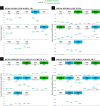
Figure 7. Statistical comparisons of muscle architectural properties between Salvator merianae (green) and Didelphis virginiana (blue) using unpaired two-sample Student’s t-tests (α = 0.05).
(A) Mean pennation angle; (B) mean normalized fascicle length; (C) mean normalized PCSA; (D) mean normalized muscle mass. Green tiles indicate Salvator is significantly greater, while blue tiles indicate Didelphis is significantly greater; white tiles reflect no significant difference. PCSA and Mm for Didelphis m. supraspinatus (SSP) and m. infraspinatus (ISP) are shown separately in gray, under m. supracoracoideus. M. infraspinatus and m. supraspinatus values for θ and Lf are too close together for differences to be visible at this scale, and are not shown in the figure. P-values shown are adjusted for multiple comparisons using the Benjamini–Hochberg procedure (also given in Table S3). Lf of m. subscapularis was significantly different prior to correction; uncorrected P-value shown in parentheses. Muscle abbreviations follow Fig. 2.
Figure 8
Figure 8. Functional morphospace comparing normalized PCSA against normalized fascicle length.
Muscles tend to vary along either one axis or the other, consistent with a tradeoff between force production (y-axis) and working range (x-axis). Muscle abbreviations and color-coding follow Fig. 2.
Figure 9
Figure 9. Predicted muscle physiological cross-sectional areas (PCSAs) for traversodontid cynodont Massetognathus pascuali (MCZVP 3691).
Salvator-like, Didelphis-like, and intermediate reconstructions are depicted with diagonally-hatched, dotted, and solid bars, respectively. Predictions are based on a reconstructed body mass of 1.437 kg. Black numbers give the value of the intermediate estimate for each muscle. Black lines show uncertainty around predictions, calculated by scaling PCSAs using the upper and lower end estimates of Massetognathus’ body mass (from percent prediction error (PPE) reported by Campione & Evans (2012)). SBC, TMJ and CBL lack error bars, as they are reconstructed from only one side of the bracket. Morphologically-informed low- and high-end estimates for the Massetognathus m. supracoracoideus are labeled as “Lo” and “Hi” respectively (see Table S5). Muscle abbreviations and color-coding follow Fig. 2. MCZVP: Museum of Comparative Zoology, Vertebrate Paleontology.

References

    1. Abdala V, Diogo R. Comparative anatomy, homologies and evolution of the pectoral and forelimb musculature of tetrapods with special attention to extant limbed amphibians and reptiles. Journal of Anatomy. 2010;217(5):536–573. doi: 10.1111/j.1469-7580.2010.01278.x. - DOI - PMC - PubMed
    1. Alexander RM, Jayes AS, Maloiy GMO, Wathuta EM. Allometry of the leg muscles of mammals. Journal of Zoology. 1981;194(4):539–552. doi: 10.1111/j.1469-7998.1981.tb04600.x. - DOI
    1. Allen V, Elsey RM, Jones N, Wright J, Hutchinson JR. Functional specialization and ontogenetic scaling of limb anatomy in Alligator mississippiensis. Journal of Anatomy. 2010;216(4):423–445. doi: 10.1111/j.1469-7580.2009.01202.x. - DOI - PMC - PubMed
    1. Allen V, Molnar J, Parker W, Pollard A, Nolan G, Hutchinson JR. Comparative architectural properties of limb muscles in Crocodylidae and Alligatoridae and their relevance to divergent use of asymmetrical gaits in extant Crocodylia. Journal of Anatomy. 2014;225(6):569–582. doi: 10.1111/joa.12245. - DOI - PMC - PubMed
    1. Ashley-Ross M. Metamorphic and speed effects on Hindlimb Kinematics during Terrestrial Locomotion in the salamander Dicamptodon Tenebrosus. Journal of Experimental Biology. 1994;193:285–305. - PubMed

LinkOut - more resources