

An Unusual Cause of Vesical Calculi
- PMID: 32117652
- PMCID: PMC7029823
- DOI: 10.7759/cureus.6701
An Unusual Cause of Vesical Calculi
Abstract
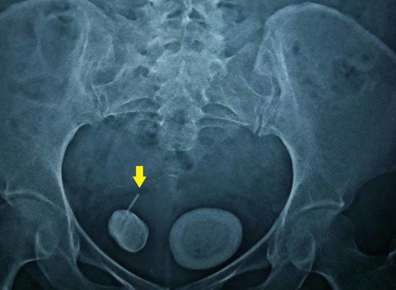
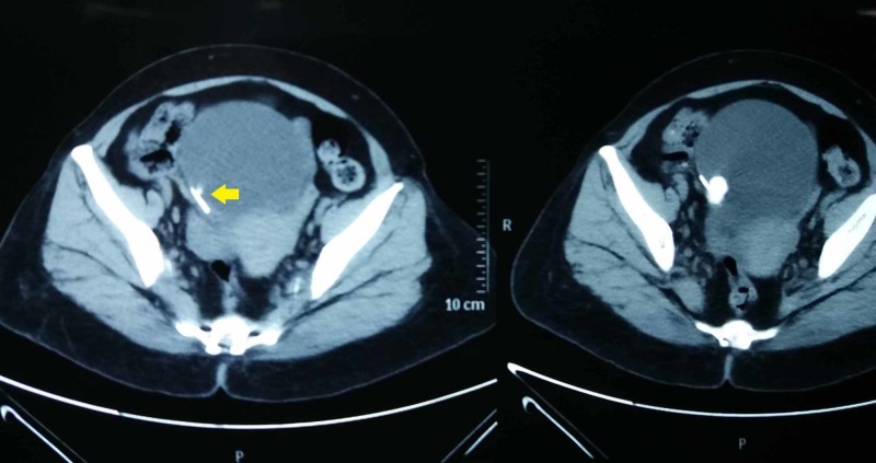
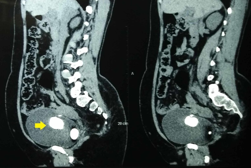
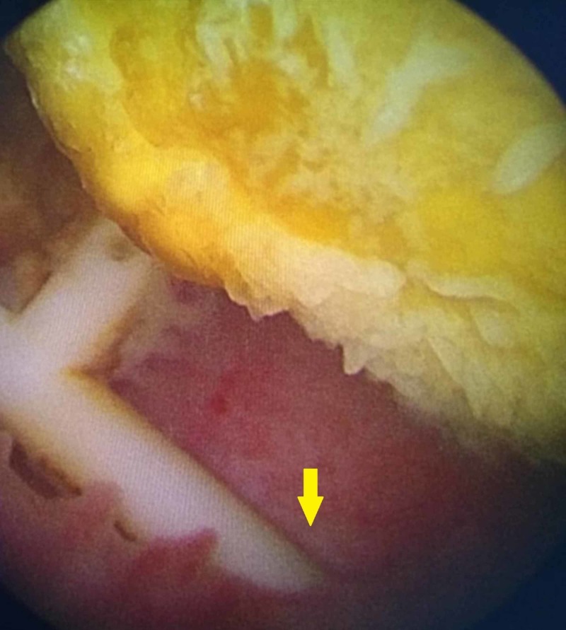
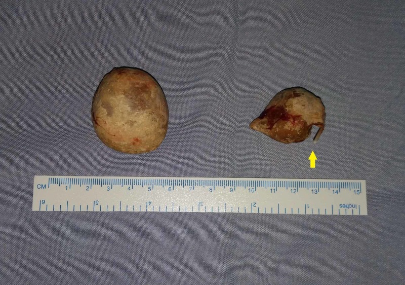
Intravesical migration is an uncommon but serious complication of intrauterine contraceptive devices. Calculus formation is common over such migrated intrauterine contraceptive devices. This dreaded complication usually presents with lower urinary tract symptoms such as suprapubic pain, frequency, and nocturia. We present a case of a 50-year-old woman with intravesical migration of copper-T device placed in the immediate postpartum period 25 years ago. She presented with dysuria, which was confirmed by computed tomography. The migrated device was encrusted with a 3.5-cm-sized stone around its vertical limb. Another stone of approximately the same size was present in the bladder. Surprisingly, the patient never had symptoms and hence she never followed up for 25 years. The stones could not be removed endoscopically, and therefore an open vesicolithotomy was performed. This case has been presented to highlight the significance of following up patients with intrauterine contraceptive devices to avoid potentially devastating complications.
Keywords: copper intrauterine contraceptive devices; encrustation; migration; vesical calculus.
Copyright © 2020, Jamburaj et al.
Conflict of interest statement
The authors have declared that no competing interests exist.
Figures
References
-
- Case of urethral foreign body: IUD perforation of the bladder with calculus formation. Gillis E, Chhiv N, Kang S, Sayegh R, Lotfipour S. https://www.ncbi.nlm.nih.gov/pubmed/20505808. Cal J Emerg Med. 2006;7:47–53. - PMC - PubMed
-
- Intrauterine device migration to the urinary bladder causing sexual dysfunction: a case report. Dimitropoulos K, Skriapas K, Karvounis G, Tzortzis V. https://www.ncbi.nlm.nih.gov/pmc/articles/PMC5074402/ Hippokratia. 2016;20:70–72. - PMC - PubMed
Publication types
LinkOut - more resources
Full Text Sources