. 2020 Feb 14:10:36.
doi: 10.3389/fonc.2020.00036. eCollection 2020.

High Expression of Integrin α3 Predicts Poor Prognosis and Promotes Tumor Metastasis and Angiogenesis by Activating the c-Src/Extracellular Signal-Regulated Protein Kinase/Focal Adhesion Kinase Signaling Pathway in Cervical Cancer

Affiliations

High Expression of Integrin α3 Predicts Poor Prognosis and Promotes Tumor Metastasis and Angiogenesis by Activating the c-Src/Extracellular Signal-Regulated Protein Kinase/Focal Adhesion Kinase Signaling Pathway in Cervical Cancer

Qiqiao Du et al. Front Oncol. .

Abstract

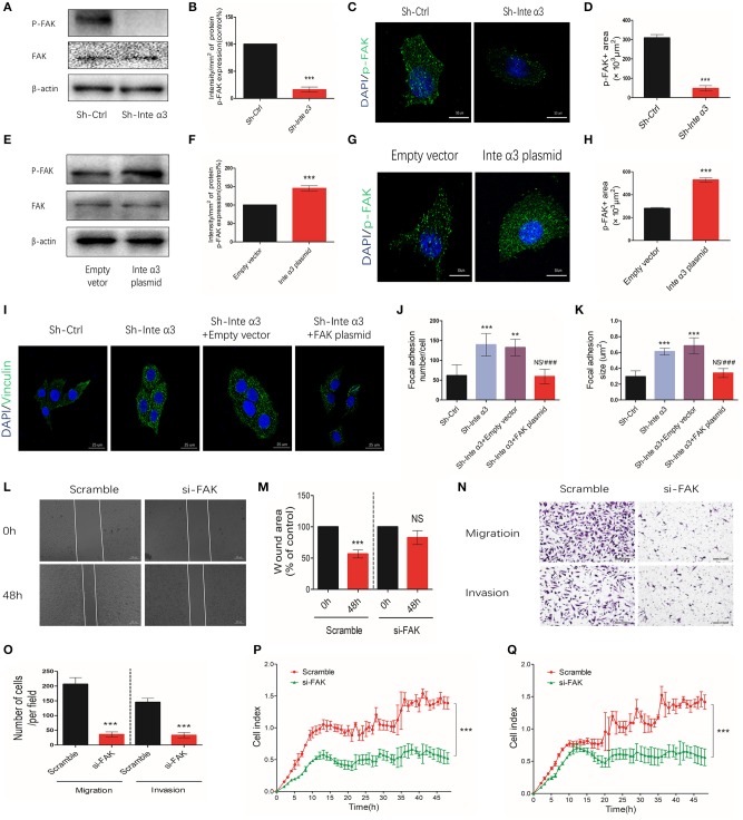
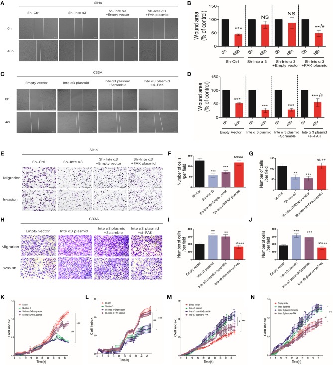
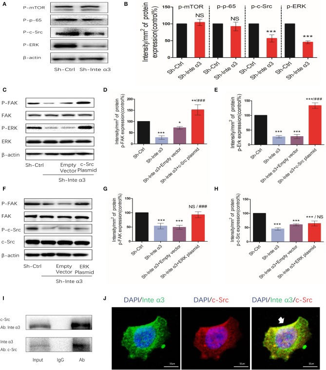
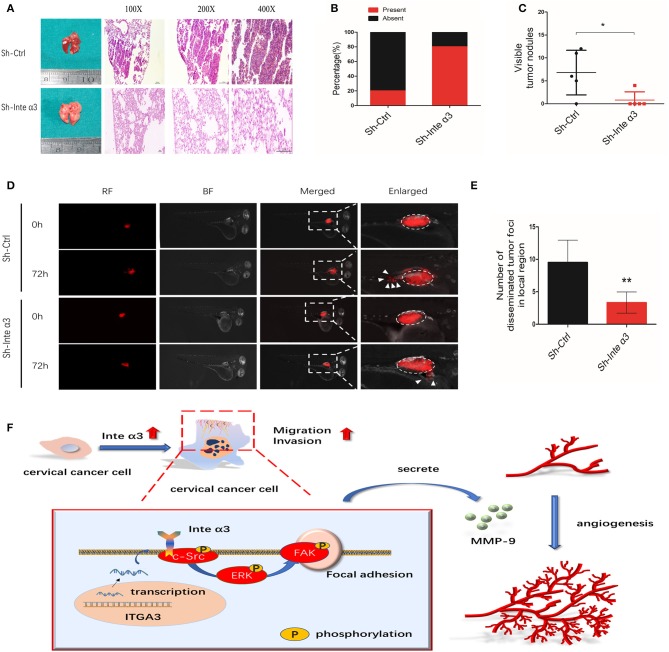
Background: Cervical cancer remains a leading cause of death in women due to metastasis to distant tissues and organs. Integrins are involved in cancer metastasis. However, whether integrin α3 participates in cervical cancer metastasis is under investigation. In this study, we explored the effect and detailed mechanism through which integrin α3 regulates cervical cell migration, invasion, and angiogenesis. Methods: First, we explored the mRNA and protein expression levels of integrin α3 in cervical cancer cell lines and tissue samples obtained from patients. After knocking down the expression of integrin α3 using shRNA, the proliferation, migration, and invasion of cervical cancer cells, as well as the possible signaling pathways involved, were investigated in vitro. In addition, tube formation, proliferation, and migration of human umbilical vein endothelial cells were tested to identify their effect on angiogenesis. Zebrafish tumor migration and nude mouse lung metastasis models were utilized for the in vivo analysis. Results: We examined samples from 142 patients with cervical cancer and 20 normal cervixes. Integrin α3 was highly expressed in patients and predicted poor overall survival and disease-free survival. In SiHa cells, treatment with integrin α3 shRNA induced the phosphorylation of protein focal adhesion kinase and enhanced focal adhesion. These events were mediated by the activation of c-Src and extracellular signal-regulated protein kinase cascades. Consequently, integrin α3 increased the migratory ability of SiHa cells. In addition, knockdown of integrin α3 decreased the tube formation, proliferation, and migration of human umbilical vein endothelial cells, as well as the levels of matrix metalloproteinase-9, indicating its effect on angiogenesis. Stable transfection with integrin α3 shRNA reduced the migratory ability of SiHa cells in the zebrafish model and diminished lung metastasis in the xenograft mouse model. Conclusion: Integrin α3 recruits the c-Src/extracellular signal-regulated protein kinase cascade, leading to phosphorylation of focal adhesion kinase. Moreover, it regulates focal adhesion, endowing cervical cancer cells with potentiated migratory and invasive ability, and promotes angiogenesis via matrix metalloproteinase-9. Our findings may shed light on the mechanism involved in cervical cancer metastasis and highlight integrin α3 as a candidate prognostic biomarker and therapeutic target in patients with cervical cancer.

Keywords: angiogenesis; cervical cancer; focal adhesion kinase; integrin α3; metastasis.


Figures

Figure 1
Figure 1
Integrin α3 is overexpressed in cervical cancer tissue and cell lines. (A) The results of the quantitative real-time (qRT)-PCR analysis showed the mRNA levels of ITGA3 expression in different cervical cancer cell lines. Normal cervix (NC) tissue was used as the normal control. (B) Western blotting was applied to show the protein levels of integrin α3 in different cervical cancer lines. (C) Quantification results of the protein intensity of (B). (D) Results of the qRT-PCR analysis of ITGA3 expression in normal cervix tissue (n = 12) and cervical cancer (CC) tissue (n = 39). (E) Western blotting was used to analyze the protein expression of integrin α3 in NC (n = 4) and CC (n = 7). (F) Quantification results of the protein intensity of (E). (G) Immunohistochemistry (IHC) assays were performed to investigate the protein expression of integrin α3 in three trial pairs of NC and CC. Original magnification: ×100. *P < 0.05 vs. corresponding control; **P < 0.01; ***P < 0.001.
Figure 2
Figure 2
Integrin α3 is frequently upregulated in cervical cancer tissue and significantly associated with overall survival and disease-free survival. (A) Representative immunohistochemistry (IHC) image of cervical cancer (CC) tissue with different staining intensities of integrin α3. (B) Representative IHC image of CC tissue of classic positive staining of integrin α3 under different magnifications (×100, ×200, and ×400). (C) Bar graph presenting the statistical composition of high/low expression in normal cervix (NC) and CC tissues. (D,E) Overall survival and disease-free survival of patients with high or low integrin α3 expression, respectively. The survival curve was constructed using the log-rank test. The P-value is shown in each panel.
Figure 3
Figure 3
Integrin α3 promotes the migration and invasion, but not proliferation, of cervical cancer cells in vitro. (A) SiHa cells were treated with control shRNA (Sh-Ctrl) and integrin α3-shRNA (Sh-Inte α3). Subsequently, immunofluorescence staining of cells with Ki-67 was performed to investigate cell proliferation. (B) Quantification of Ki-67-positive cells per field in (A) (original magnification: ×100). (C) The MTT assay was used to analyze the proliferation rate between the Sh-Ctrl and Sh-Inte α3 groups. (D) The wound healing assay was applied in SiHa cells to explore the effect of integrin α3 on cell migration in vitro in the Sh-Ctrl and Sh-Inte α3 groups, respectively. (E) Quantification of the wound area in (D). (F) The wound healing assay was applied in C33A cells to explore the effect of integrin α3 on cell migration in vitro in the empty vector and Inte α3 plasmid groups. (G) Quantification of the wound area in (F). (H–K) Transwell migration and invasion assays were performed to investigate the effect of integrin α3 on the migration and invasion of cervical cancer cells either knocking down in SiHa or overexpressing in C33A. Quantification results of the migration and invasion of cells per field in (I,K). (L,M) Migration and invasion were tested in the Sh-Ctrl and Sh-Inte α3 groups through real-time migration/invasion monitoring. (N,O) Migration and invasion were tested in empty vector and Inte α3 plasmid groups through real-time migration/invasion monitoring. All experiments were repeated thrice with consistent results, and the representative images are shown. *P < 0.05 vs. corresponding control; **P < 0.01; ***P < 0.001; ##P < 0.01.
Figure 4
Figure 4
Integrin α3 impairs focal adhesion formation by activating focal adhesion kinase (FAK). (A,B) Western blotting results of phosphor-Y397-FAK (p-FAK) after knocking down the expression of integrin α3 using shRNA in vitro and quantification of the intensity of p-FAK protein. (C,D) After knocking down integrin α3, immunofluorescence was applied to detect the expression levels of p-FAK and quantification of the p-FAK-positive area. (E,F) Western blotting results of p-FAK after overexpressing of integrin α3 and quantification of the intensity of p-FAK protein. (G,H) Immunofluorescence results of p-FAK after overexpressing integrin α3 and quantification of the p-FAK-positive area. (I) Focal adhesion formation was investigated through vinculin immunofluorescence in the following four groups: Sh-Ctrl, Sh-Inte α3, Sh-Inte α+empty vector, and Sh-Inte α3+FAK plasmid. (J,K) Quantification of focal adhesion (number and size) in (I). (L) The wound healing assay was applied to explore the migration of cervical cancer cells in Scramble and si-FAK groups, and the quantification is presented in (M). (N) Transwell migration and invasion assays were performed in the two groups. (O) Quantification of migration and invasion (cell number) per field. (P,Q) Results related to cell migration and invasion using real-time migration/invasion monitoring. All experiments were repeated thrice with consistent results, and the representative images are shown. **P < 0.01; ***P < 0.001; ###P < 0.001 vs. Sh-Inte α3+empty vector.
Figure 5
Figure 5
Integrin α3 promotes the migration and invasion of cells via focal adhesion kinase (FAK). (A) The wound healing assay was applied in SiHa cell to explore the migration of cervical cancer cells in the four groups—Sh-Ctrl, Sh-Inte α3, Sh-Inte α+empty vector, and Sh-Inte α3+FAK plasmid—and the quantification is presented in (B). (C) The wound healing assay was applied in C33A cell in the four groups—empty vector, Inte α3 plasmid, Inte α3 plasmid+Scramble, and Inte α3 plasmid+si-FAK—and the quantification is presented in (D). (E) Transwell migration and invasion assays in SiHa cells were performed in the four groups in (A), and corresponding quantification of migration and invasion cells is shown in (F,G). (H) Transwell migration and invasion assays in C33A cells were performed in the four groups in (C), and corresponding quantification of migration and invasion cells is shown in (I,J). (K,L) Results related to cell migration and invasion using real-time migration/invasion monitoring in SiHa cell in the aforementioned groups in (A). (M,N) Results related to cell migration and invasion using real-time migration/invasion monitoring in C33A cell in the aforementioned groups in (C). **P < 0.01; ***P < 0.001; #P < 0.05 vs. corresponding group (e.g., Sh-Inte α+empty vector vs. Sh-Inte α3+FAK plasmid); ##P < 0.01; ###P < 0.01.
Figure 6
Figure 6
Integrin α3 activates focal adhesion kinase (FAK) via the c-Src/extracellular signal-regulated protein kinase (ERK) signaling pathway. (A) Phosphorylated mammalian target of rapamycin (mTOR), p-65, c-Src, and ERK were examined through western blotting in the Sh-Ctrl and Sh-Inte α groups; the corresponding quantification of the protein intensity is shown in (B). (C) The levels of phosphorylated FAK and ERK, as well as total FAK, ERK, and β-actin, were determined via western blotting among the Sh-Ctrl, Sh-Inte α3, Sh-Inte α3+empty vector, and Sh-Inte α3+c-Src plasmid groups; the corresponding quantification of p-FAK and p-c-Src protein intensity is presented in (D,E). (F) Among the Sh-Ctrl, Sh-Inte α3, Sh-Inte α3+empty vector, and Sh-Inte α3+ERK plasmid groups, the levels of phosphorylated FAK and c-Src, as well as total FAK, c-Src, and β-actin, were examined through western blotting; the quantification of p-FAK and p-ERK protein intensity is shown in (G,H). (I) Representative image of co-immunoprecipitation using anti-c-Src or anti-integrin α3, and subsequent detection of the other protein. (J) Representative immunofluorescence images showing colocalizations between integrin α3 and c-Src. Nuclei were stained with 4′-6-diamidino-2-phenylindole (DAPI) (blue), anti-c-Src linked to Alexa Fluor (red), anti-integrin α3 linked to fluorescein isothiocyanate (FITC) (green); original magnification: ×200. All experiments were repeated thrice with consistent results, and the representative images are shown. *P < 0.05 vs. control group; **P < 0.01; ***P < 0.001; ###P < 0.01.
Figure 7
Figure 7
Integrin α3 promotes angiogenesis by secreting metalloproteinase-9 (MMP-9) in vitro. (A,B) Immunohistochemistry (IHC) staining and corresponding quantification of CD34 in the high/low integrin α3 expression groups in cervical cancer (CC) tissue. Original magnification: ×100, ×200, and ×400. (C) Correlation of IHC score and microvascular density. The correlation coefficient is shown in the panel. (D) The human umbilical vein endothelial cell (HUVEC) tube formation assay was applied to explore the effect of integrin α3 on angiogenesis. (E) Quantification of the total tube length per field is shown in (D). (F,G) Real-time migration/invasion monitoring was used for the detection of migration and invasion of HUVECs treated with the cell culture medium from the Sh-Ctrl and Sh-Inte α3 groups. (H,I) ELISA for MMP-9 was performed after knocking down integrin α3 and focal adhesion kinase (FAK). All experiments were repeated thrice with consistent results, and the representative images are shown. *P < 0.05 vs. control group; ***P < 0.001.
Figure 8
Figure 8
Integrin α3 promotes metastasis in vivo; a proposed schematic representation of the mechanism. (A) Typical H&E images of pulmonary metastatic foci in the Sh-Ctrl group and normal pulmonary tissues in the Sh-Inte α3 group are shown. Original magnification: ×100, ×200, and ×400. (B) Percentages of mice with or without metastatic foci in their lungs in the aforementioned two groups (n = 5). (C) Visible tumor nodules in the lungs were calculated in these two groups. (D) The zebrafish tumor migration model was applied to explore the migration ability of cells in these two groups. (E) Quantification of the number of disseminated foci in the local region of (D). (F) Proposed schematic representation of the mechanism demonstrating that integrin α3 activates the c-Src/Erk/focal adhesion kinase (FAK) signaling pathway and promotes cervical cancer metastasis and angiogenesis. *P < 0.05 vs. control group; **P < 0.01.

Similar articles

Cited by

References

    1. Bray F, Ferlay J, Soerjomataram I, Siegel RL, Torre LA, Jemal A. Global cancer statistics 2018: GLOBOCAN estimates of incidence and mortality worldwide for 36 cancers in 185 countries. CA Cancer J Clin. (2018) 68:394–424. 10.3322/caac.21492 - DOI - PubMed
    1. Campbell CMP, Menezes LJ, Paskett ED, Giuliano AR. Prevention of invasive cervical cancer in the United States: past, present, and future. Cancer Epidemiol Biomarkers Prev. (2012) 21:1402–8. 10.1158/1055-9965.EPI-11-1158 - DOI - PMC - PubMed
    1. Qureshi R, Arora H, Rizvi MA. EMT in cervical cancer: its role in tumour progression and response to therapy. Cancer Lett. (2015) 356:321–31. 10.1016/j.canlet.2014.09.021 - DOI - PubMed
    1. Wan L, Pantel K, Kang Y. Tumor metastasis: moving new biological insights into the clinic. Nat Med. (2013) 19:1450–64. 10.1038/nm.3391 - DOI - PubMed
    1. Vicente-Manzanares M, Sanchez-Madrid F. Targeting the integrin interactome in human disease. Curr Opin Cell Biol. (2018) 55:17–23. 10.1016/j.ceb.2018.05.010 - DOI - PubMed