

Magnetic Resonance vs. Intraoral Ultrasonography in the Preoperative Assessment of Oral Squamous Cell Carcinoma: A Systematic Review and Meta-Analysis
- PMID: 32117789
- PMCID: PMC7010633
- DOI: 10.3389/fonc.2019.01571
Magnetic Resonance vs. Intraoral Ultrasonography in the Preoperative Assessment of Oral Squamous Cell Carcinoma: A Systematic Review and Meta-Analysis
Abstract
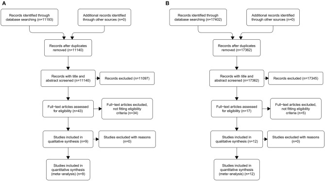
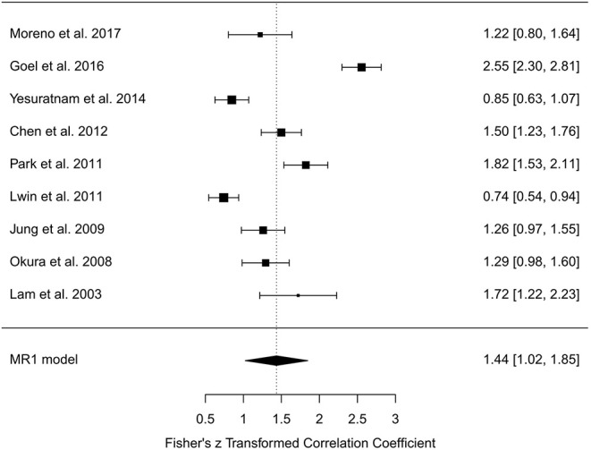
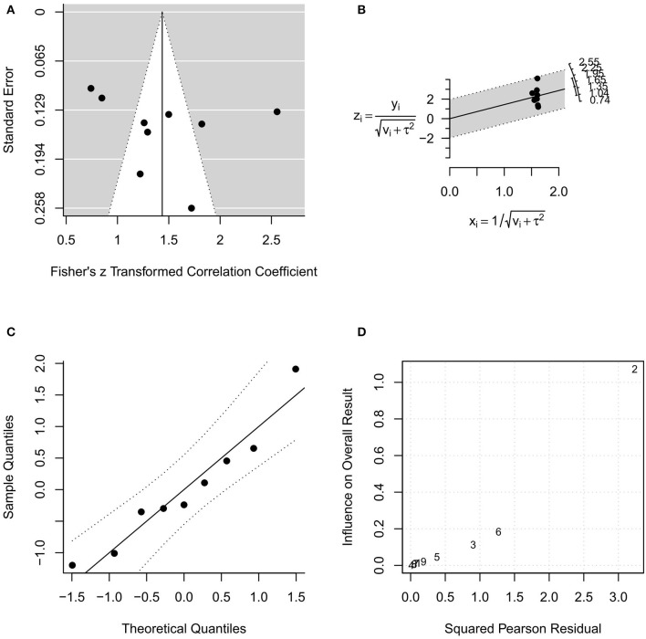
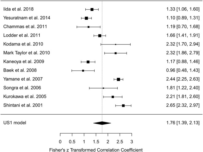
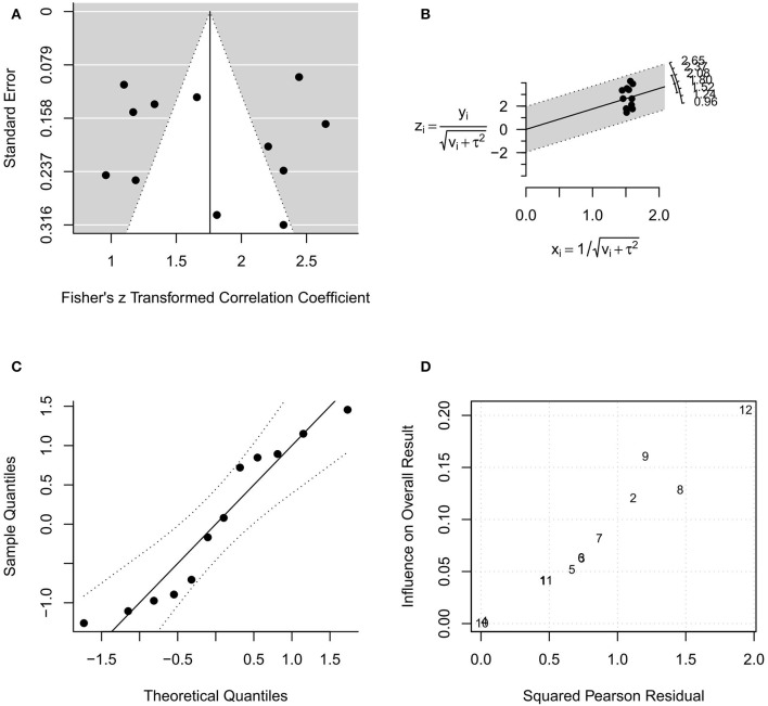
Background: Preoperative assessment is critical to decide the most adequate surgical strategy for oral squamous cell carcinoma (SCC). Magnetic resonance (MR) and intraoral ultrasonography (US) have been reported to be of great value for preoperative estimation of depth of invasion (DOI) and/or tumor thickness (TT). This review aims to analyze the accuracy of MR and intraoral US in determining DOI/TT in oral SCC, by assuming histological evaluation as the reference method. Methods: The procedure was conducted following the modified 2009 Preferred Reporting Items for Systematic Reviews and Meta-Analyses (PRISMA) statement. We performed a systematic search of papers on PubMed, Scopus, Web of Science, and Cochrane Library databases until July 31st, 2019. For quantitative synthesis, we included nine studies (487 patients) focused on MR, and 12 (520 patients) focused on intraoral US. The Pearson correlation coefficient (r) between DOI/TT evaluated with MR or intraoral US was assumed as effect size. A meta-analysis (MA) for each study group (MR and US) was performed by using the random-effects models with the DerSimonian-Laird estimator and r-to-z transformation. Results: In the MA for MR studies, a high heterogeneity was found (I 2 = 94.84%; Q = 154.915, P < 0.001). No significant risk of bias occurred by evaluating funnel plot asymmetry (P = 0.563). The pooled (overall) r of the MR studies was 0.87 (95% CI from 0.82 to 0.92), whereas the pooled r-to-z transformed was 1.44 (95% CI from 1.02 to 1.85). In the MA for US studies a high heterogeneity was found (I 2 = 93.56%; Q = 170.884, P < 0.001). However, no significant risk of bias occurred (P = 0.779). The pooled r of the US studies was 0.96 (95% CI from 0.94 to 0.97), whereas the pooled r-to-z transformed was 1.76 (95% CI from 1.39 to 2.13). These outputs were confirmed in additional MA performed by enrolling only MR (n = 8) and US (n = 11) studies that evaluated TT. Conclusions: MR and intraoral US seem to be promising approaches for preoperative assessment of DOI/TT in oral SCC. Remarkably, a higher pooled r and r-to-z transformed were observed in the intraoral US studies, suggesting that this approach could be more closely related to histopathological findings.
Keywords: depth of invasion (DOI); magnetic resonance imaging (MRI); oral cavity; squamous cell cancer (SCC); tumor thickness; ultrasound.
Copyright © 2020 Marchi, Filauro, Iandelli, Carobbio, Mazzola, Santori, Parrinello, Canevari, Piazza and Peretti.
Figures
References
-
- Edge SB, Byrd DR, Compton CC, Fritz AG, Green FL, Trotti A. editors. AJCC Cancer Staging Manual. 7th ed New York, NY: Dordrecht; Heidelberg; London: Springer; (2010).
Publication types
LinkOut - more resources
Full Text Sources
Research Materials