

Reconstruction of Large Skeletal Defects: Current Clinical Therapeutic Strategies and Future Directions Using 3D Printing
- PMID: 32117940
- PMCID: PMC7029716
- DOI: 10.3389/fbioe.2020.00061
Reconstruction of Large Skeletal Defects: Current Clinical Therapeutic Strategies and Future Directions Using 3D Printing
Abstract
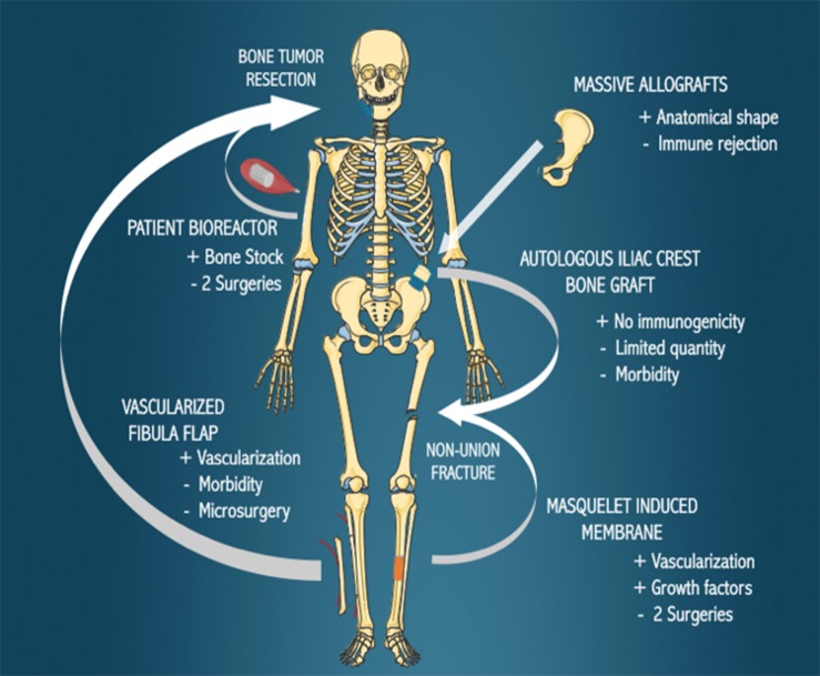
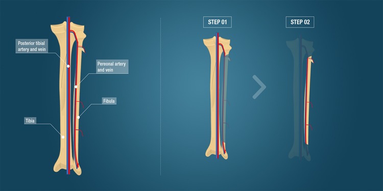
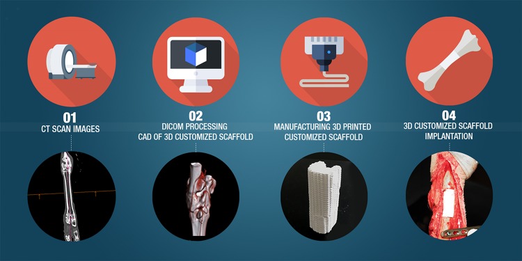
The healing of bone fractures is a well-orchestrated physiological process involving multiple cell types and signaling molecules interacting at the fracture site to replace and repair bone tissue without scar formation. However, when the lesion is too large, normal healing is compromised. These so-called non-union bone fractures, mostly arising due to trauma, tumor resection or disease, represent a major therapeutic challenge for orthopedic and reconstructive surgeons. In this review, we firstly present the current commonly employed surgical strategies comprising auto-, allo-, and xenograft transplantations, as well as synthetic biomaterials. Further to this, we discuss the multiple factors influencing the effectiveness of the reconstructive therapy. One essential parameter is adequate vascularization that ensures the vitality of the bone grafts thereby supporting the regeneration process, however deficient vascularization presents a frequently encountered problem in current management strategies. To address this challenge, vascularized bone grafts, including free or pedicled fibula flaps, or in situ approaches using the Masquelet induced membrane, or the patient's body as a bioreactor, comprise feasible alternatives. Finally, we highlight future directions and novel strategies such as 3D printing and bioprinting which could overcome some of the current challenges in the field of bone defect reconstruction, with the benefit of fabricating personalized and vascularized scaffolds.
Keywords: bone regeneration; large bone defects; three-dimensional printing; tissue engineering; vascularization.
Copyright © 2020 Vidal, Kampleitner, Brennan, Hoornaert and Layrolle.
Figures
References
-
- Aronson J., Harrison B. H., Stewart C. L., Harp J. H. (1989). The histology of distraction osteogenesis using different external fixators. Clin. Orthop. Relat. Res. 241 106–116. - PubMed
-
- Athanasiou V. T., Papachristou D. J., Panagopoulos A., Saridis A., Scopa C. D., Megas P. (2010). Histological comparison of autograft, allograft-DBM, xenograft, and synthetic grafts in a trabecular bone defect: an experimental study in rabbits. Med. Sci. Monit. 16 BR24–BR31. - PubMed
Publication types
LinkOut - more resources
Full Text Sources
Other Literature Sources