Review
. 2020 Feb 11:8:17.
doi: 10.3389/fcell.2020.00017. eCollection 2020.

T Cell Dysfunction and Exhaustion in Cancer

Affiliations
Review

T Cell Dysfunction and Exhaustion in Cancer

Zhen Zhang et al. Front Cell Dev Biol. .

Abstract

Tumor immunotherapy is a promising therapeutic strategy for patients with advanced cancers. T cells are key mediators of antitumor function that specifically recognize and react to tumor-expressing antigens and have proven critical for cancer immunotherapy. However, T cells are not as effective against cancer as expected. This is partly because T cells enter a dysfunctional or exhausted state, which is characterized by sustained expression of inhibitory receptors and a transcriptional state distinct from that of functional effector or memory T cells. T cell dysfunction induces the out of control of tumors. Recently, T cell dysfunction has been investigated in many experimental and clinical settings. The molecular definition of T cell dysfunction and the underlying causes of the T cell dysfunction has been advanced regardless of the fact that the pathways involved are not well elucidated, which proposing promising therapeutic opportunities in clinic. In this review, we will discuss the recent advances in the molecular mechanisms that affect TME and induce T cell dysfunction, and the development of promising immunotherapies to counteract the mechanisms of tumor-induced T cell dysfunction. Better understanding these underlying mechanisms may lead to new strategies to improve the clinical outcome of patients with cancer.

Keywords: T cells dysfunction; cancer immunotherapy; extrinsic factors; intrinsic factors; tumor microenvironment.


Figures

FIGURE 1
FIGURE 1
The intrinsic factors regulating T cell dysfunction. In response to T cell receptors (TCRs), co-stimulatory and growth factor cytokines activate PI3K/Akt/mTOR signaling pathways, which induce glucose transporter-1 (Glut-1) expression and enhance T cell proliferation and cytokine production. Activation of mTOR leads to the expression of downstream transcriptional regulators such as HIF-1α and c-Myc. However, an increased AMP to ATP ratio activates AMP-activated protein kinase (AMPK), which in turn inhibits mTOR activity and enhances fatty acid oxidation, which maintains long term T-cell survival and formation of memory T cells. The Transcription factors such as HIF-1α, NR4A1, TOX, Eomes, T-bet, Blimp-1, NFAT and BATF regulate PD-1 expression and have been implicated in T cell exhaustion and dysfunction.
FIGURE 2
FIGURE 2
Immunosuppressive cells or factors have been implicated in CD8+ T cell dysfunction in TME. The ARG1, iNOS, TGF-β and ROS are secreted by MDSCs or TAMs and induce CD8+ T cell dysfunction. Both MDSCs and tumor cells may suppress CD8+ T cell proliferation through IDO hydrolyzation of tryptophan in the presence of IFN-γ. Kynurenine inhibits CD8+ T cell activation. MDSCs may additionally produce immunosuppressive cytokines like IL-10, TGF-β and induce Tregs. The upregulation of PD-L1 on MDSCs, TAMs and tumor cells induced CD8+ T cell exhaustion by binding to PD-1 on T cells. Tumor cells also express CD39 and CD73 on their surface, facilitating the metabolism of extracellular ATP into AMP and finally into adenosine, which induce CD8+ T cell dysfunction. CAFs are involved in impairing anti-tumor T cell responses by secreting chemokines such as CXCL1 and CXCL2 to tumors and polarizing them toward the M2 phenotype. Furthermore, the expression of PD-L2 or FASL on CAFs bind to corresponding PD-1 and FAS receptors, respectively, causing CD8+ T cell dysfunction.
FIGURE 3
FIGURE 3
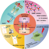
Combinatorial therapeutic strategies to reverse the T cell dysfunction. A growing number of studies propose to evaluate the efficacy of immune checkpoint blockade antibodies together with: (a) CAR-T cell therapy; (b) inhibitors of soluble mediators targeting IL-10, TGF-β, DNA methyltransferase and histone deacetylase; (c) CSF1R antibody targeting activating receptors on TAMs to inhibit the polarization of TAM; and (d) neoantigen vaccines.

References

    1. Ahmed N., Brawley V., Hegde M., Bielamowicz K., Kalra M., Landi D., et al. (2017). HER2-specific chimeric antigen receptor-modified virus-specific T cells for progressive glioblastoma: a phase 1 dose-escalation trial. JAMA Oncol. 3 1094–1101. 10.1001/jamaoncol.2017.0184 - DOI - PMC - PubMed
    1. Alfei F., Kanev K., Hofmann M., Wu M., Ghoneim H. E., Roelli P., et al. (2019). TOX reinforces the phenotype and longevity of exhausted T cells in chronic viral infection. Nature 571 265–269. 10.1038/s41586-019-1326-9 - DOI - PubMed
    1. Ambade A., Satishchandran A., Saha B., Gyongyosi B., Lowe P., Kodys K., et al. (2016). Hepatocellular carcinoma is accelerated by NASH involving M2 macrophage polarization mediated by hif-1alphainduced IL-10. Oncoimmunology 5:e1221557. 10.1080/2162402x.2016.1221557 - DOI - PMC - PubMed
    1. Bally A. P., Austin J. W., Boss J. M. (2016). Genetic and epigenetic regulation of PD-1 expression. J. Immunol. 196 2431–2437. 10.4049/jimmunol.1502643 - DOI - PMC - PubMed
    1. Benner B., Scarberry L., Suarez-Kelly L. P., Duggan M. C., Campbell A. R., Smith E., et al. (2019). Generation of monocyte-derived tumor-associated macrophages using tumor-conditioned media provides a novel method to study tumor-associated macrophages in vitro. J. Immunother. Cancer 7:140. 10.1186/s40425-019-0622-0 - DOI - PMC - PubMed

LinkOut - more resources