Review
. 2020 Feb 7:8:63.
doi: 10.3389/fcell.2020.00063. eCollection 2020.

CDP-Diacylglycerol Synthases (CDS): Gateway to Phosphatidylinositol and Cardiolipin Synthesis

Affiliations
Review

CDP-Diacylglycerol Synthases (CDS): Gateway to Phosphatidylinositol and Cardiolipin Synthesis

Nicholas J Blunsom et al. Front Cell Dev Biol. .

Abstract

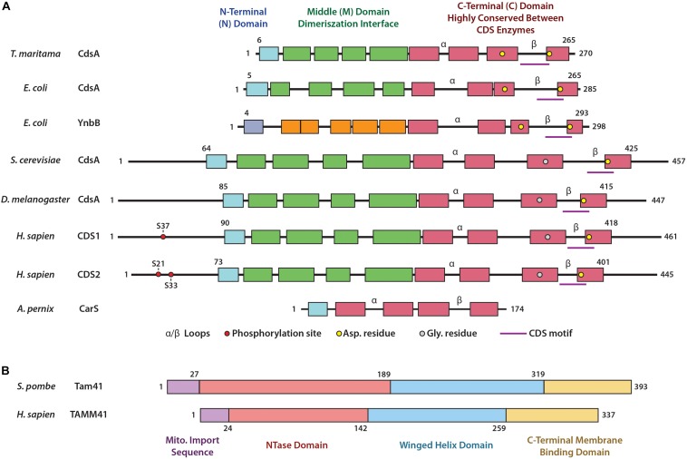
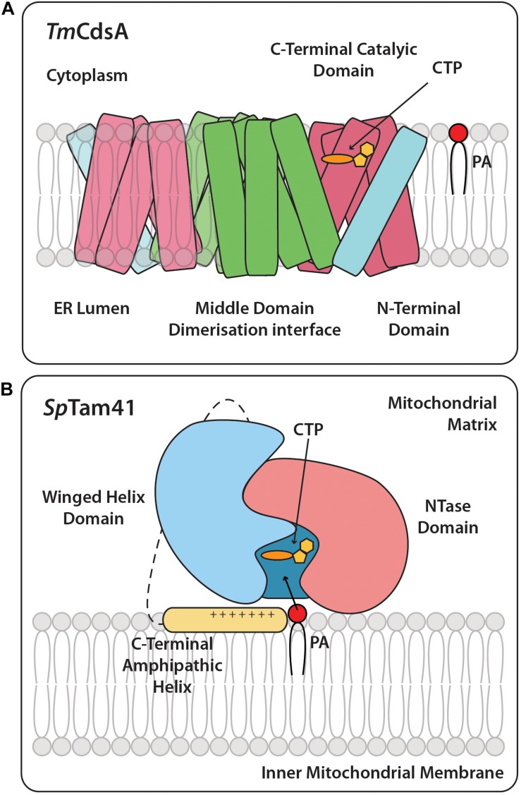
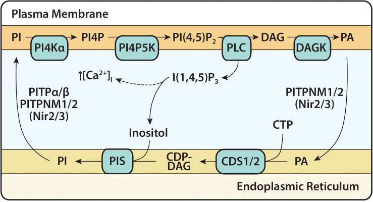
Cytidine diphosphate diacylglycerol (CDP-DAG) is a key intermediate in the synthesis of phosphatidylinositol (PI) and cardiolipin (CL). Both PI and CL have highly specialized roles in cells. PI can be phosphorylated and these phosphorylated derivatives play major roles in signal transduction, membrane traffic, and maintenance of the actin cytoskeletal network. CL is the signature lipid of mitochondria and has a plethora of functions including maintenance of cristae morphology, mitochondrial fission, and fusion and for electron transport chain super complex formation. Both lipids are synthesized in different organelles although they share the common intermediate, CDP-DAG. CDP-DAG is synthesized from phosphatidic acid (PA) and CTP by enzymes that display CDP-DAG synthase activities. Two families of enzymes, CDS and TAMM41, which bear no sequence or structural relationship, have now been identified. TAMM41 is a peripheral membrane protein localized in the inner mitochondrial membrane required for CL synthesis. CDS enzymes are ancient integral membrane proteins found in all three domains of life. In mammals, they provide CDP-DAG for PI synthesis and for phosphatidylglycerol (PG) and CL synthesis in prokaryotes. CDS enzymes are critical for maintaining phosphoinositide levels during phospholipase C (PLC) signaling. Hydrolysis of PI (4,5) bisphosphate by PLC requires the resynthesis of PI and CDS enzymes catalyze the rate-limiting step in the process. In mammals, the protein products of two CDS genes (CDS1 and CDS2) localize to the ER and it is suggested that CDS2 is the major CDS for this process. Expression of CDS enzymes are regulated by transcription factors and CDS enzymes may also contribute to CL synthesis in mitochondria. Studies of CDS enzymes in protozoa reveal spatial segregation of CDS enzymes from the rest of the machinery required for both PI and CL synthesis identifying a key gap in our understanding of how CDP-DAG can cross the different membrane compartments in protozoa and in mammals.

Keywords: CDP-diacylglycerol; TAMM41; endoplasmic reticulum; lipid synthesis; mitochondria; phosphatidic acid; phosphatidylinositol; phospholipase C.


Figures

FIGURE 1
FIGURE 1
Synthesis of phosphatidylinositol (PI) at the ER and of cardiolipin (CL) in mitochondria. PA, in the ER, is converted to CDP-DAG via the CDS enzymes. CDP-DAG is used by PIS, to synthesize PI. PA can be transferred from the ER to mitochondria, where it is transported across the OMM and IMS to the IMM. Here, TAMM41 utilizes PA to make CDP-DAG, then PGP via PGPS, and PG via PTPMT1. Finally, CL is made by CLS using PG and another molecule of CDP-DAG. It could be the case the CDP-DAG is transported across the cytosol from the ER to mitochondria, where it can be utilized for CL synthesis as discussed in the text. PA, phosphatidic acid; PIS, PI synthase; TAMM41, enzyme with CDS activity; PGPS, phosphatidylglycerolphosphate synthase; CLS, cardiolipin synthase; PI, phosphatidylinositol; CDP-DAG, CDP-diacylglycerol; PGP, phosphatidylglycerol phosphate; PTPMT1, protein tyrosine phosphatase mitochondrial 1; PG, phosphatidylglycerol; CL, cardiolipin; MAM, mitochondrial associated membranes; OMM, outer mitochondrial membrane; IMS, inner mitochondrial space; IMM, inner mitochondrial membrane.
FIGURE 2
FIGURE 2
Domain structures of CDS enzymes and of Tam41. (A) Domain structures of the CDS enzymes based upon the bacterial enzyme TmCdsA from T. maritama. These include the N-terminal domain (blue), the Middle domain involved in dimerization (green) and the highly conserved catalytic C-terminal domain (red). Within the C-terminal domain lies the α and β loops, the CDS motif, and residues involved in coordination of metal ions crucial for catalysis. Phosphorylation sites of HsCDS1/2 are also included. (B) Domain structures of TAMM41 enzymes based upon the yeast enzyme SpTam41 from S. pombe: these include the mitochondrial import sequence (purple), the NTase domain (orange) and winged helix domain (blue) (which form the catalytic binding pocket), and the C-terminal membrane binding domain (yellow).
FIGURE 3
FIGURE 3
Cartoon structures of TmCdsA and SpTam41. (A) CDS is an integral membrane protein and is present as a dimer and accepts the two substrates, CTP, and PA. On the cytoplasmic side of each TmCdsA monomer, a funnel-shaped cavity indents half way into the membrane region. The cavity has two wide openings, which enable it to receive dual substrates, CTP from the cytoplasm and PA from the lipid bilayer at the same time. The conversion of CTP and PA into CDP-DAG and pyrophosphate occurs through a process involving the transfer of CMP group from CTP onto the phosphate group of PA. Adapted from Liu X. et al. (2014). Color coding is the same as TmCdsA in Figure 2. (B) The full length SpTam41 exists as a monomer and is associated to the matrix side of the inner mitochondrial membrane by the membrane binding domain at the C-terminal region (colored yellow). CTP and PA sequentially bind to the active site of Tam41. The enzymatic conversion of CTP and PA into CDP-DAG and pyrophosphate occurs through a process involving the transfer of CMP group from CTP onto the phosphate group of PA. Adapted from Jiao et al. (2019). Color coding is the same as SpTam41 in Figure 2.
FIGURE 4
FIGURE 4
The PI(4,5)P2 cycle. The PI(4,5)P2 cycle begins with the hydrolysis of PI(4,5)P2 and formation of the second messengers, I(1,4,5)P3 and DAG. I(1,4,5)P3 causes an increase in intracellular calcium concentration before being dephosphorylated into inositol. DAG is phosphorylated to PA at the plasma membrane (PM) by DAG kinase (DAGK) and transferred to the ER via Class IIa PITPs (PI/PA transfer proteins) PITPNM1/2. At the ER, PA, and CTP are converted to CDP-DAG by CDS enzymes (CDS1 and CDS2). CDP-DAG is synthesized into PI and this is catalyzed by the enzyme PI synthase (PIS). PI is transferred to the PM by Class I PITPs (PI/PC transfer proteins) PITPα/β and Class IIa PITPNM1/2, for phosphorylation to PI(4,5)P2 by the resident enzymes, PI4KIIIα and PIP5K. PITPNM1/2 are also known as Nir2/Nir3.

References

    1. Acehan D., Malhotra A., Xu Y., Ren M., Stokes D. L., Schlame M. (2011). Cardiolipin affects the supramolecular organization of ATP synthase in mitochondria. Biophys. J. 100 2184–2192. 10.1016/j.bpj.2011.03.031 - DOI - PMC - PubMed
    1. AhYoung A. P., Jiang J., Zhang J., Khoi Dang X., Loo J. A., Zhou Z. H. (2015). Conserved SMP domains of the ERMES complex bind phospholipids and mediate tether assembly. Proc. Natl. Acad. Sci. U.S.A. 112 E3179–E3188. 10.1073/pnas.1422363112 - DOI - PMC - PubMed
    1. Alfonso Pecchio A. R., Cardozo Gizzi A. M., Renner M. L., Molina-Calavita M., Caputto B. L. (2011). c-Fos activates and physically interacts with specific enzymes of the pathway of synthesis of polyphosphoinositides. Mol. Biol. Cell 22 4716–4725. 10.1091/mbc.E11-03-0259 - DOI - PMC - PubMed
    1. Alhouayek M., Masquelier J., Muccioli G. G. (2018). Lysophosphatidylinositols, from cell membrane constituents to GPR55 Ligands. Trends Pharmacol. Sci. 39 586–604. 10.1016/j.tips.2018.02.011 - DOI - PubMed
    1. Anderson K. E., Kielkowska A., Durrant T. N., Juvin V., Clark J., Stephens L. R., et al. (2013). Lysophosphatidylinositol-acyltransferase-1 (LPIAT1) is required to maintain physiological levels of PtdIns and PtdInsP(2) in the mouse. PLoS. One 8:e58425. 10.1371/journal.pone.0058425 - DOI - PMC - PubMed