. 2020 Feb 5:7:45.
doi: 10.3389/fvets.2020.00045. eCollection 2020.

In-vivo Activity of IFN-λ and IFN-α Against Bovine-Viral-Diarrhea Virus in a Mouse Model

Affiliations

In-vivo Activity of IFN-λ and IFN-α Against Bovine-Viral-Diarrhea Virus in a Mouse Model

María Eugenia Quintana et al. Front Vet Sci. .

Abstract

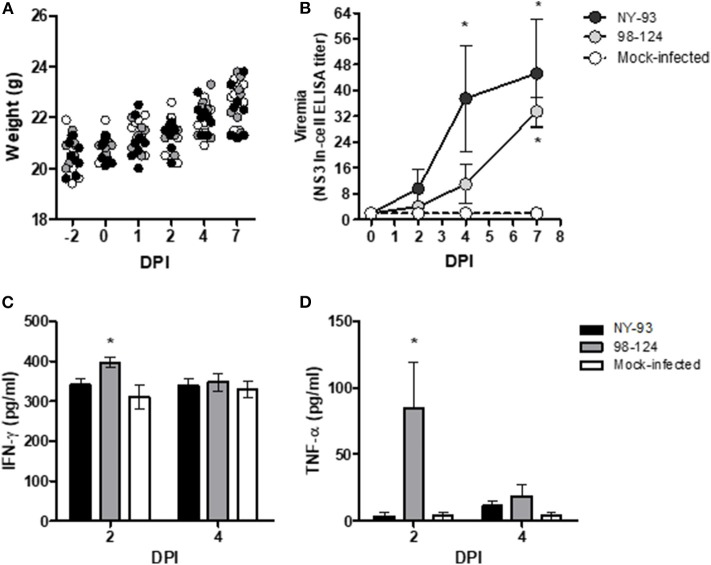
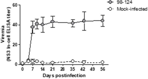
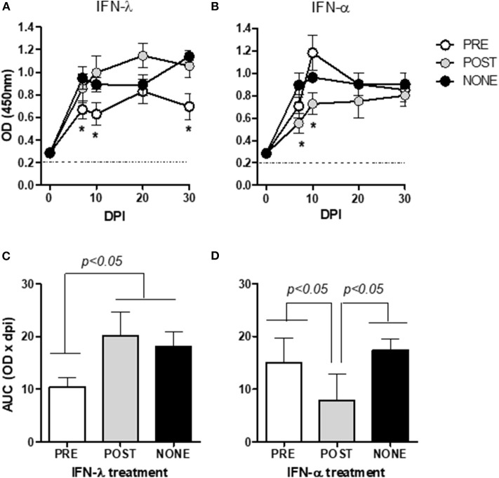
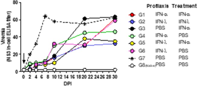
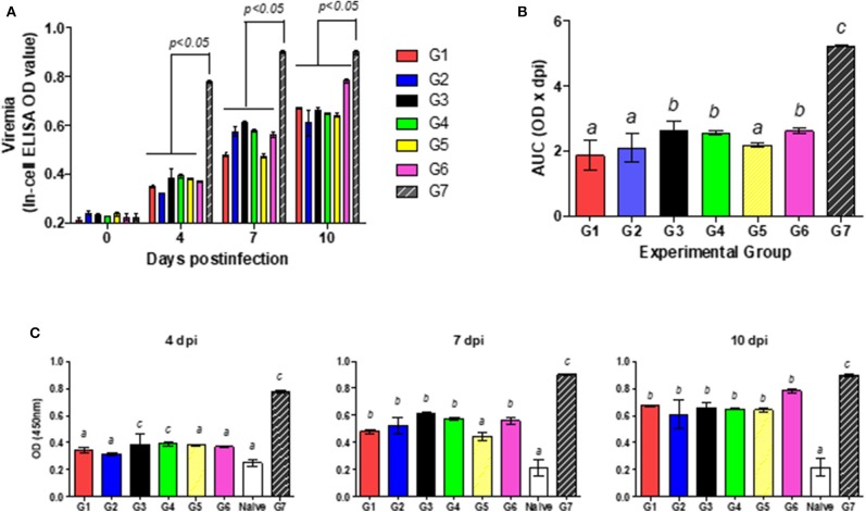
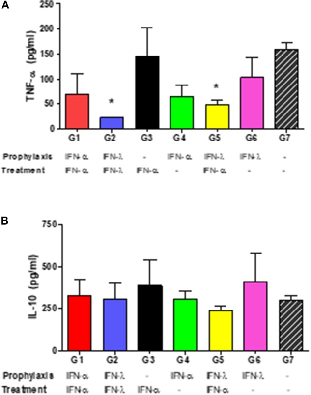
Bovine-viral-diarrhea virus (BVDV) can cause significant economic losses in livestock. The disease is controlled with vaccination and bovines are susceptible until vaccine immunity develops and may remain vulnerable if a persistently infected animal is left on the farm; therefore, an antiviral agent that reduces virus infectivity can be a useful tool in control programs. Although many compounds with promising in-vitro efficacy have been identified, the lack of laboratory-animal models limited their potential for further clinical development. Recently, we described the activity of type I and III interferons, IFN-α and IFN-λ respectively, against several BVDV strains in-vitro. In this study, we analyzed the in-vivo efficacy of both IFNs using a BALB/c-mouse model. Mice infected with two type-2 BVDV field strains developed a viremia with different kinetics, depending on the infecting strain's virulence, that persisted for 56 days post-infection (dpi). Mice infected with the low-virulence strain elicited high systemic TNF-α levels at 2 dpi. IFNs were first applied subcutaneously 1 day before or after infection. The two IFNs reduced viremia with different kinetics, depending on whether either one was applied before or after infection. In a second experiment, we increased the number of applications of both IFNs. All the treatments reduced viremia compared to untreated mice. The application of IFN-λ pre- and post-infection reduced viremia over time. This study is the first proof of the concept of the antiviral potency of IFN-λ against BVDV in-vivo, thus encouraging further trails for a potential use of this cytokine in cattle.

Keywords: antiviral activity; bovine-viral-diarrhea virus; interferon-α; interferon-λ; mouse model.


Figures

Figure 1
Figure 1
Mouse-BVDV–infection model. BALB/c mice were infected with two ncp BVDV genotype-2 strains, 98-124 and NY-93. In the figure, body weight (A); viremia (B); and the levels of the cytokines IFN-γ (C) and TNF-α (D) in pg/ml are plotted as functions of the time in days post-infection (DPI). Key to the point and bar textures: black, primary antibody against the NY-93 BVDV strain; gray, primary antibody against the 98-124 BVDV strain; white, mock-infected mice. The asterisks (*) denote values significantly higher than those measured in the mock-infected mice (p < 0.05).
Figure 2
Figure 2
Kinetics of viral replication, as measured in the BVDV levels of blood samples of mice infected with the BVDV 98-124 strain by NS3-In-cell ELISA. Samples were taken at different time points from 0 to 56 days post-infection. Values are expressed as the mean NS3 titers ± SD of triplicates.
Figure 3
Figure 3
Effect of IFN prophylaxis or treatment in viremia. In the figures, the kinetics of viral replication, are expressed as mean OD450 ± SD of triplicate values. Mice were inoculated with either IFN-λ (A) or IFN-α (B) before (PRE, white circles) or after (POST, gray circles) infection, or infected but left untreated (NONE, black circles). The figures of (C) (IFN-λ) and (D) (IFN-α) are plots of the areas under the respective curves from (A,B) (mean ± SD) for the three inoculation protocols (PRE, POST, and NONE). The corresponding values for the noninfected mice (mock-infected groups) were below the detection levels of the assay and are thus not included in the figures. The asterisks (*) in (A,B) denote values significantly lower than those measured in the NONE group (p < 0.05). The dotted horizontal line in figures (A,B) depict the cut-off value. DPI, days post-infection.
Figure 4
Figure 4
Effect of combined treatment in viremia. In the figure, the kinetics of viral replication in pools of serum, expressed as NS3 titers, is plotted as a function of the days post-infection (DPI). The arrow marks the time of infection. The key to the experimental groups to the right of the figure, summarizes the IFN-pre- and/or –post-infection protocol for each of the experimental groups (G1–G8).
Figure 5
Figure 5
Effect of combined treatment in viremia. (A) Viral replication was measured as the mean OD450 levels ± the SD reflecting the NS3 titer, for each of the experimental groups G1–G7. (B) The mean value ± the SD for the area under the curve (AUC) resulting from the bar heights in (A) from days 1 through 10 is plotted for each of the experimental groups G1–G7. (C) Viral replication was measured as in (A) for each individual animal in the groups G1–G7 plus Group G8 (the naïve mice) at Day 4 (left figure), Day 7 (middle figure), and Day 10 (right figure) post-infection. In (B,C), different letters above the bars mark significant differences between groups (p < 0.05).
Figure 6
Figure 6
Systemic cytokine levels. In the figures, the mean concentrations ± SD of IFN-α (A) or IL-10 (B) in pg/ml of individual serum samples at 2 dpi are plotted for each experimental group. The asterisks (*) denote significant differences compared to the corresponding levels measured in the infected, untreated animals (Group 7) at a p < 0.05.

References

    1. Becher P, Orlich M, Shannon AD, Horner G, König M, Thiel HJ. Phylogenetic analysis of pestiviruses from domestic and wild ruminants. J Gen Virol. (1997) 78:1357–66. 10.1099/0022-1317-78-6-1357 - DOI - PubMed
    1. Terpstra C, Wensvoort G. A congenital persistent infection of bovine virus diarrhoea virus in pigs: clinical, virological and immunological observations. Vet Q. (1997) 19:97–101. 10.1080/01652176.1997.9694750 - DOI - PubMed
    1. Lee KM, Gillespie JH. Propagation of virus diarrhea virus of cattle in tissue culture. Am J Vet Res. (1957) 18:952–3. - PubMed
    1. New York State College of Veterinary Medicine New York State Veterinary College (1911). The Cornell Veterinarian. Ithaca, NY: Published under the auspices of the Alumni Association and Society of Comparative Medicine, New York State Veterinary College, Cornell University.
    1. Lazear HM, Daniels BP, Pinto AK, Huang AC, Vick SC, Doyle SE, et al. . Interferon-λ restricts west nile virus neuroinvasion by tightening the blood-brain barrier. Sci Transl Med. (2015) 7:284ra59. 10.1126/scitranslmed.aaa4304 - DOI - PMC - PubMed

LinkOut - more resources