. 2020 Feb 17;5(7):3258-3270.
doi: 10.1021/acsomega.9b03307. eCollection 2020 Feb 25.

In Vitro and Ex Vivo Efficacy of Novel Trp-Arg Rich Analogue of α-MSH against Staphylococcus aureus

Affiliations

In Vitro and Ex Vivo Efficacy of Novel Trp-Arg Rich Analogue of α-MSH against Staphylococcus aureus

Jyotsna Singh et al. ACS Omega. .

Abstract

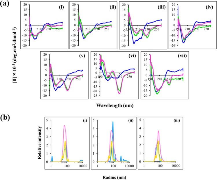
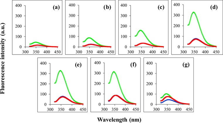
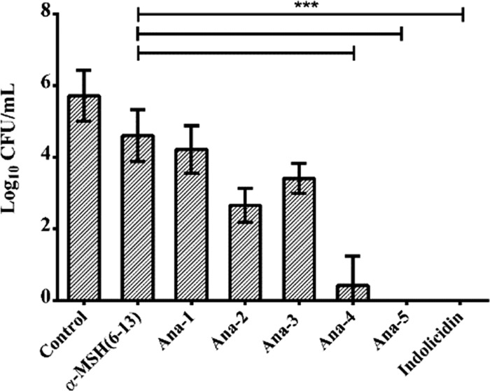
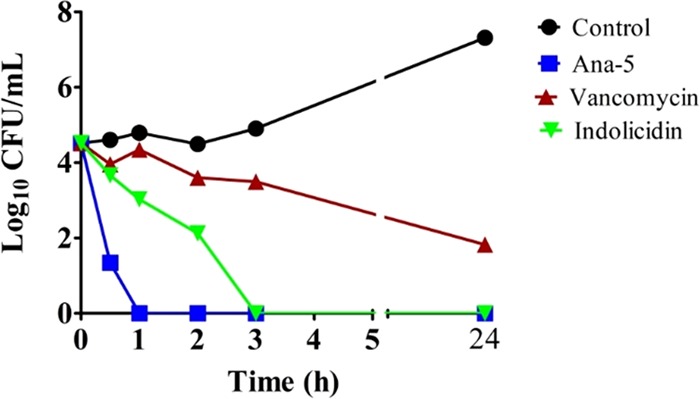
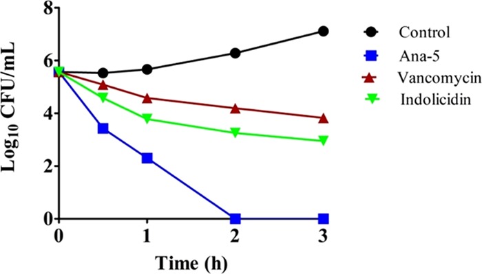
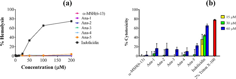
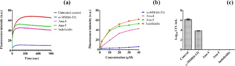
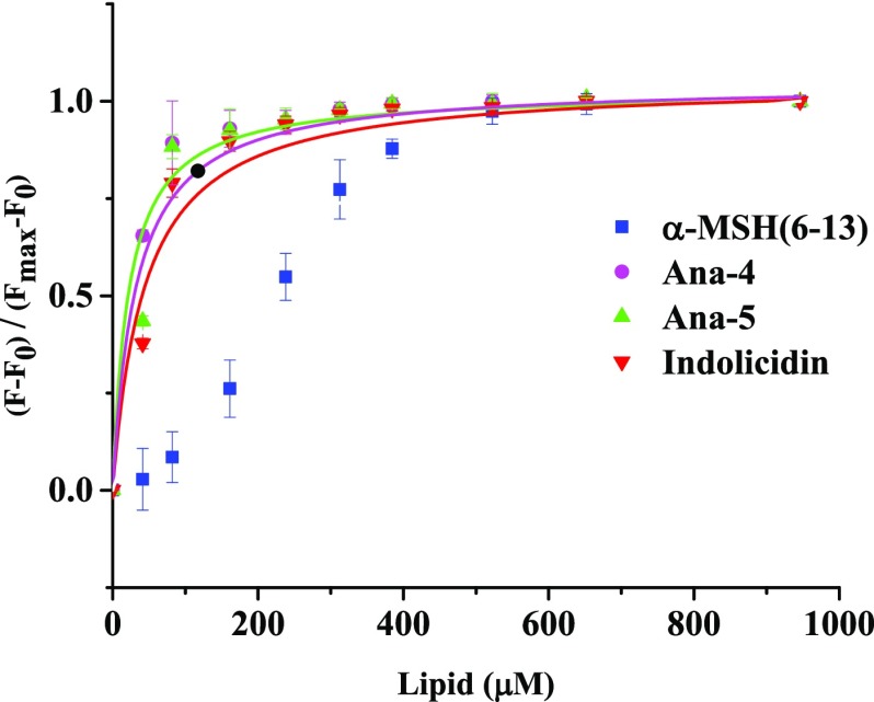
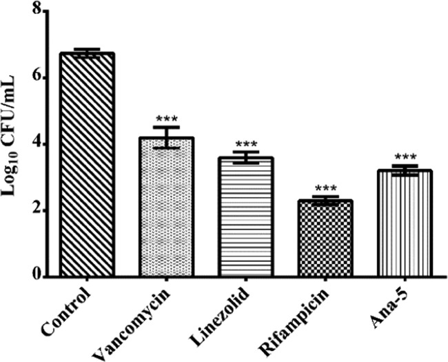
Antimicrobial peptides (AMPs), an essential component of innate immunity, are very important resources for human therapeutics to counter the current threat of drug resistance. We have previously established that one such AMP, α-melanocyte stimulating hormone (α-MSH), an endogenous neuropeptide, and its derivatives have potent antimicrobial activity against Staphylococcus aureus, including methicillin-resistant S. aureus (MRSA). However, the immense potential of α-MSH for therapeutic development against staphylococcal infections is marred by its reduced efficacy in the presence of standard microbiological culture medium. To overcome this issue, in this study, we designed a series of five novel analogues of the C-terminal fragment of α-MSH, i.e., α-MSH(6-13), by replacing uncharged and less hydrophobic residues with tryptophan and arginine to increase the hydrophobicity and cationic charge of the peptide, respectively. While all of the peptides showed a preferential interaction with negatively charged phospholipid vesicles, the most hydrophobic and cationic peptide, i.e., Ana-5, exhibited the highest activity against S. aureus cells while maintaining cell selectivity. Moreover, Ana-5 could retain its activity even in complex media like the Mueller Hinton broth and displayed rapid bactericidal activity in the presence of serum. Ana-5 also caused rapid bacterial membrane depolarization, permeabilization, and cell lysis and was able to bind to polyanionic plasmid DNA suggesting a possible dual mode of action of the peptide. Importantly, Ana-5 was able to eradicate intracellular S. aureus in fibroblast cells similar to conventional antibiotics. Collectively, in the present study, we obtained a potent α-MSH-based analogue with excellent staphylocidal potency in microbial growth medium and ex vivo efficacy, which may translate into therapeutic application.


Conflict of interest statement

The authors declare no competing financial interest.

Figures

Figure 1
Figure 1
Structural characterization of the designed peptides. (a) Circular dichroism spectra of α-MSH(6–13) and its analogues in 5 mM PB (blue), DMPC/DMPG (7:3, w/w) SUVs (green), and DMPC/DMPG (1:1, w/w) SUVs (pink). (i) α-MSH(6–13), (ii) Ana-1, (iii) Ana-2, (iv) Ana-3, (v) Ana-4, (vi) Ana-5, and (vii) indolicidin, at peptide and lipid concentrations of 35 and 1453 μM, respectively (L/P molar ratio, 41.5:1). The spectrum of each peptide represents an average of three scans and was plotted as mean residue ellipticity (in deg cm2 dmol–1) against wavelength (nm). (b) Dynamic light scattering profile expressed as relative intensity vs radius (nm) for DMPC/DMPG (1:1, w/w) SUVs alone (yellow), peptides dissolved in water (blue), and with added DMPC/DMPG (1:1, w/w) SUVs (pink). Size distribution of the peptides (i) Ana-2, (ii) Ana-4, and (iii) Ana-5, at peptide and lipid concentrations of 35 and 1453 μM, respectively (L/P molar ratio of 41.5:1). Experiments were performed twice, and representative spectra are shown here.
Figure 2
Figure 2
Tryptophan fluorescence emission spectra of peptides in different milieus: in buffer (blue), DMPC (red), and DMPC/DMPG (7:3, w/w) (green) SUVs. (a) α-MSH(6–13), (b) Ana-1, (c) Ana-2, (d) Ana-3, (e) Ana-4, (f) Ana-5, and (g) indolicidin. Peptide and lipid concentrations were 14.5 and 726 μM, respectively, i.e., L/P molar ratio was 50:1.
Figure 3
Figure 3
Antibacterial activity of α-MSH(6–13) and designed analogues against the mid-log phase of S. aureus. Cell viability was observed after 2 h incubation with 10 μM α-MSH(6–13) and its analogues in 5 mM N-(2-hydroxyethyl)piperazine-N′-ethanesulfonic acid (HEPES)-20 mM glucose buffer, pH 7.4. Experiments were done on three different days and presented as mean ± standard deviation (SD) (***P < 0.001).
Figure 4
Figure 4
In vitro bacterial killing kinetics of log phase cells of S. aureus in culture medium. The cells were treated with Ana-5, vancomycin, and indolicidin at their 4 × MIC in MHB media for 24 h. The experiments were repeated on three different days, and similar data were obtained. Representative data are shown here.
Figure 5
Figure 5
Antibacterial activity against S. aureus in the presence of 10% serum. Log phase bacterial cells were treated with Ana-5, vancomycin, and indolicidin at their 4 × MIC in MHB media with 10% serum for 3 h. The experiments were repeated on three different days, and similar data were obtained. Representative data are shown here.
Figure 6
Figure 6
Toxicity studies of α-MSH(6–13) and its analogues against mammalian cells. (a) % Hemolysis of mouse RBCs upon 1 h treatment with different concentrations of the peptides. (b) % Cytotoxicity observed in 3T3 murine fibroblast cell line on treatment with the peptides at 15, 30, and 60 μM concentrations for 2 h. Each assay was done in triplicate on two different days.
Figure 7
Figure 7
Membrane depolarization efficacy of the peptides. (a) Depolarization kinetics measured at a fixed concentration of 30 μM of α-MSH(6–13), Ana-4, Ana-5, and indolicidin for 15 min. (b) Concentration-dependent effect of the peptides on the membrane potential of S. aureus measured by fluorescence intensity of the dye. (c) Corresponding cell viability of the DiSC3(5)-loaded cells after immediate exposure to 30 μM of the peptides in 5 mM HEPES-20 mM glucose buffer, pH 7.4.
Figure 8
Figure 8
Binding isotherms derived by plotting fractions of the membrane-bound peptide (FF0)/(FmaxF0) as a function of the concentration of the lipid. The Trp fluorescence intensity at 295 nm was recorded by titrating 10 μM of the peptides with increasing molar concentrations of SUVs in 10 mM TES buffer (pH 7.4). Data were fitted (OriginLab Corp.) with the hyperbolic saturation curve for DMPC/DMPG (7:3, w/w) in the case of Ana-4 (pink, ●), Ana-5 (green, ▲), and indolicidin (red, ▼), whereas for the parent peptide α-MSH(6–13) (blue, ■), due to weak initial binding, the data points could not be fitted with the same. The data acquired are presented (mean ± SD) after background subtraction and dilution correction.
Figure 9
Figure 9
S. aureus membrane perturbation by Ana-5. Membrane permeabilization measured by the percentage of calcein leakage from calcein-AM-loaded 106 CFU mL–1 bacterial cells after 2 h treatment with α-MSH(6–13) and Ana-5. (a) Histograms (from left to right) for calcein-loaded mid-log phase cells that were (i) untreated, (ii) treated with α-MSH(6–13), and (iii) treated with Ana-5. (b) % Calcein leakage from S. aureus cells after treatment with α-MSH(6–13) and Ana-5 at 30 μM concentration. (c) Corresponding % survival of cells after treatment with the same concentration of peptides for 2 h (**P < 0.01, ***P < 0.001). (d) Scanning electron micrographs of S. aureus: (i) untreated control, treated with 50 μM of (ii) α-MSH(6–13) and (iii) Ana-5 at 50 000× magnification.
Figure 10
Figure 10
Binding of Ana-4, Ana-5, and indolicidin with plasmid DNA assessed through gel retardation assay. Peptide interaction with plasmid DNA (pBluescriptII SK(+)) was determined via inhibition of DNA migration in 1% (w/v) agarose gel. Different peptide concentrations were incubated with 100 ng of plasmid DNA for 1 h. Lane 1 contains plasmid DNA, and lanes 2–5, 6–9, and 10–13 contain Ana-4, Ana-5, and indolicidin, respectively. The four lanes of each peptide are in decreasing order of peptide concentration, i.e., 100, 50, 25, and 12.5 μM, respectively.
Figure 11
Figure 11
Ex vivo efficacy of Ana-5, vancomycin, linezolid, and rifampicin at their 5 × MIC in a coculture model of S. aureus and 3T3 murine fibroblast cell line at MOI 10:1. The log reduction of intracellular S. aureus cells upon 24 h incubation with Ana-5 and the antibiotics is shown here. Data were acquired on three different days in duplicates and represent mean ± SD (***P < 0.001).

References

    1. Tong S. Y. C.; Davis J. S.; Eichenberger E.; Holland T. L.; Fowler V. G. Staphylococcus aureus infections: epidemiology, pathophysiology, clinical manifestations, and management. Clin. Microbiol. Rev. 2015, 28, 603–661. 10.1128/CMR.00134-14. - DOI - PMC - PubMed
    1. Thomer L.; Schneewind O.; Missiakas D. Pathogenesis of Staphylococcus aureus bloodstream infections. Annu. Rev. Pathol.: Mech. Dis. 2016, 11, 343–364. 10.1146/annurev-pathol-012615-044351. - DOI - PMC - PubMed
    1. Foster T. J.; Geoghegan J. A.; Ganesh V. K.; Höök M. Adhesion, invasion and evasion: the many functions of the surface proteins of Staphylococcus aureus. Nat. Rev. Microbiol. 2014, 12, 49.10.1038/nrmicro3161. - DOI - PMC - PubMed
    1. Mahlapuu M.; Håkansson J.; Ringstad L.; Björn C. Antimicrobial peptides: an emerging category of therapeutic agents. Front. Cell. Infect. Microbiol. 2016, 6, 194.10.3389/fcimb.2016.00194. - DOI - PMC - PubMed
    1. Gonzalez-Rey E.; Ganea D.; Delgado M. Neuropeptides: keeping the balance between pathogen immunity and immune tolerance. Curr. Opin. Pharmacol. 2010, 10, 473–481. 10.1016/j.coph.2010.03.003. - DOI - PMC - PubMed

LinkOut - more resources