

Noninvasive quantification of SIRT1 expression-activity and pharmacologic inhibition in a rat model of intracerebral glioma using 2-[18F]BzAHA PET/CT/MRI
- PMID: 32118205
- PMCID: PMC7034639
- DOI: 10.1093/noajnl/vdaa006
Noninvasive quantification of SIRT1 expression-activity and pharmacologic inhibition in a rat model of intracerebral glioma using 2-[18F]BzAHA PET/CT/MRI
Abstract
Background: Several studies demonstrated that glioblastoma multiforme progression and recurrence is linked to epigenetic regulatory mechanisms. Sirtuin 1 (SIRT1) plays an important role in glioma progression, invasion, and treatment response and is a potential therapeutic target. The aim of this study is to test the feasibility of 2-[18F]BzAHA for quantitative imaging of SIRT1 expression-activity and monitoring pharmacologic inhibition in a rat model of intracerebral glioma.
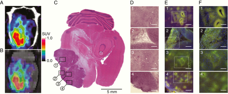
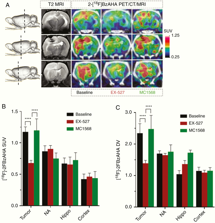
Methods: Sprague Dawley rats bearing 9L (N = 12) intracerebral gliomas were injected with 2-[18F]BzAHA (300-500 µCi/animal i.v.) and dynamic positron-emission tomography (PET) imaging was performed for 60 min. Then, SIRT1 expression in 9L tumors (N = 6) was studied by immunofluorescence microscopy (IF). Two days later, rats with 9L gliomas were treated either with SIRT1 specific inhibitor EX-527 (5 mg/kg, i.p.; N = 3) or with histone deacetylases class IIa specific inhibitor MC1568 (30 mg/kg, i.p.; N = 3) and 30 min later were injected i.v. with 2-[18F]BzAHA. PET-computerized tomography-magnetic resonance (PET/CT/MR) images acquired after EX-527 and MC1568 treatments were co-registered with baseline images.
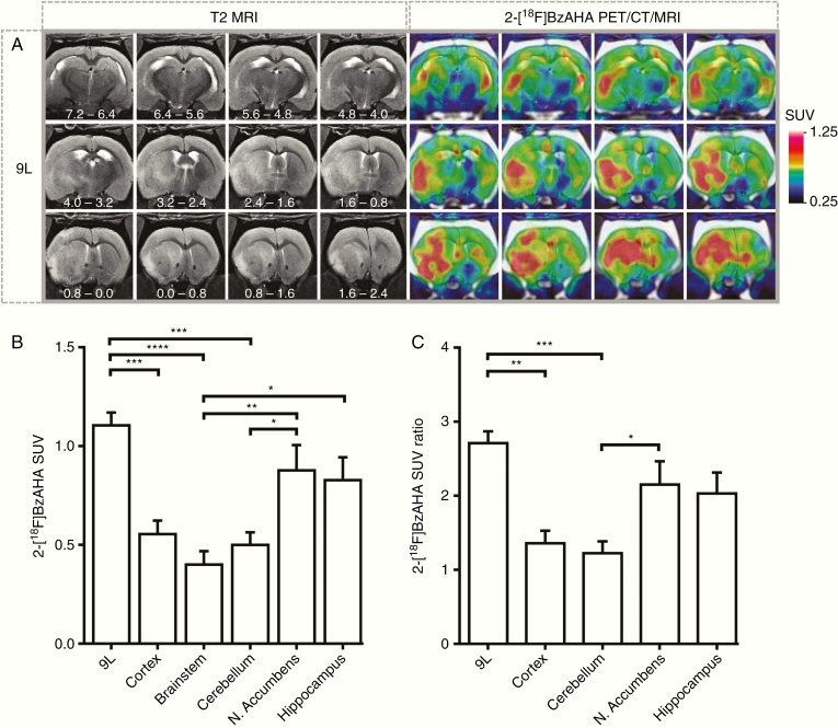
Results: Standard uptake values (SUVs) of 2-[18F]BzAHA in 9L tumors measured at 20 min post-radiotracer administration were 1.11 ± 0.058 and had a tumor-to-brainstem SUV ratio of 2.73 ± 0.141. IF of 9L gliomas revealed heterogeneous upregulation of SIRT1, especially in hypoxic and peri-necrotic regions. Significant reduction in 2-[18F]BzAHA SUV and distribution volume in 9L tumors was observed after administration of EX-527, but not MC1568.
Conclusions: PET/CT/MRI with 2-[18F]BzAHA can facilitate studies to elucidate the roles of SIRT1 in gliomagenesis and progression, as well as to optimize therapeutic doses of novel SIRT1 inhibitors.
Keywords: EX-527; SIRT1; epigenetics; glioma; molecular imaging.
© The Author(s) 2020. Published by Oxford University Press, the Society for Neuro-Oncology and the European Association of Neuro-Oncology.
Figures
References
-
- Kreth S, Thon N, Kreth FW. Epigenetics in human gliomas. Cancer Lett. 2014;342(2):185–192. - PubMed
-
- Portela A, Esteller M. Epigenetic modifications and human disease. Nat Biotechnol. 2010;28(10):1057–1068. - PubMed
-
- Berdasco M, Esteller M. Aberrant epigenetic landscape in cancer: how cellular identity goes awry. Dev Cell. 2010;19(5):698–711. - PubMed
Grants and funding
LinkOut - more resources
Full Text Sources
Miscellaneous