

Genetic Basis of Extramedullary Plasmablastic Transformation of Multiple Myeloma
- PMID: 32118627
- PMCID: PMC7225029
- DOI: 10.1097/PAS.0000000000001459
Genetic Basis of Extramedullary Plasmablastic Transformation of Multiple Myeloma
Abstract
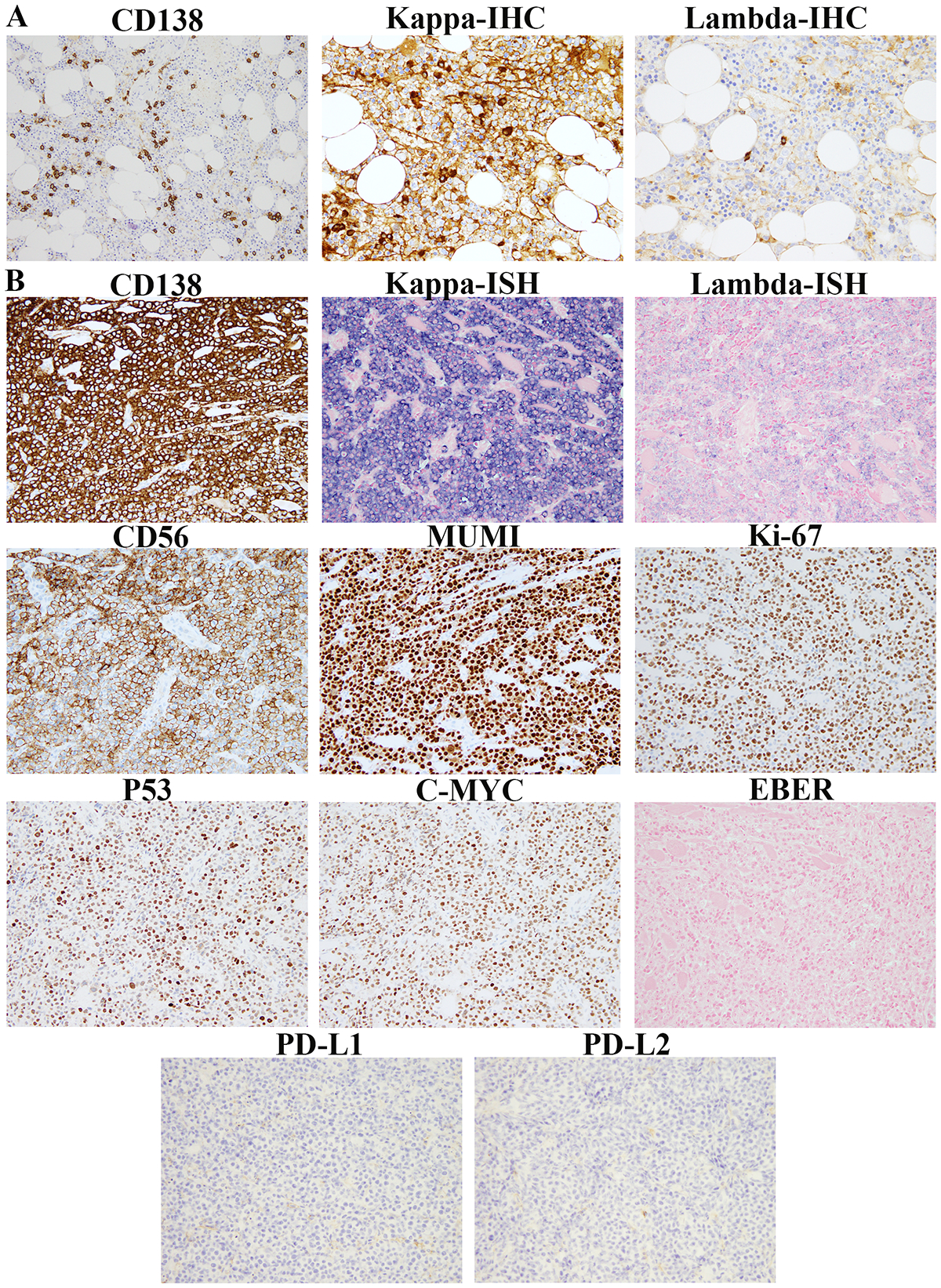
In patients with multiple myeloma, plasmablastic transformation in the bone marrow is rare and associated with poor outcomes. The significance of discordant extramedullary plasmablastic transformation in patients with small, mature clonal plasma cells in the bone marrow has not been well studied. Here, we report the clinicopathologic, cytogenetic, and molecular features of 10 such patients (male/female: 6/4, median age: 65 y, range: 48 to 76 y) with an established diagnosis of multiple myeloma in the bone marrow composed of small, mature plasma cells in parallel with a concurrent or subsequent extramedullary plasmablastic transformation. Eight patients with available survival data showed an overall aggressive clinical course with a median survival of 4.5 months after the diagnosis of extramedullary plasmablastic transformation, despite aggressive treatment and even in patients with low-level bone marrow involvement. Pathologically, the extramedullary plasmablastic myeloma were clonally related to the corresponding bone marrow plasma cells, showed high levels of CMYC and/or P53 expression with a high Ki-67 proliferation index by immunohistochemistry and harbored more complex genomic aberrations including frequent mutations in the RAS pathway and MYC rearrangements compared with their bone marrow counterparts. In summary, although genetic and immunohistochemical studies were not uniformly performed on all cases due to the retrospective nature of this study, our data suggest that discordant extramedullary plasmablastic transformation of multiple myeloma has an aggressive clinical course and is characterized by frequent mutations in the RAS pathway and more complex genomic abnormalities.
Conflict of interest statement
Conflict-of-interest disclosure: A.D. has received consultancy fees from Roche, Corvus Pharmaceuticals, Physicians’ Education Resource, Seattle Genetics, Peerview Institute, Oncology Specialty Group, Pharmacyclics, Celgene, Novartis, Takeda, EUSAPharma and research grants from Roche. W.X. has received research support from Stemline Therapeutics. The other authors have nothing to disclose.
Figures


References
-
- Swerdlow SCE, Harris NE, et al. WHO classification of tumours of haematopoietic and lymphoid tissue.. Lyon, IARC press;. 2017.
-
- Lorsbach RB, Hsi ED, Dogan A, et al. Plasma cell myeloma and related neoplasms. American journal of clinical pathology. 2011;136:168–182. - PubMed
-
- Bartl R, Frisch B, Burkhardt R, et al. Bone marrow histology in myeloma: its importance in diagnosis, prognosis, classification and staging. Br J Haematol. 1982;51:361–375. - PubMed
-
- Bartl R, Frisch B, Fateh-Moghadam A, et al. Histologic classification and staging of multiple myeloma. A retrospective and prospective study of 674 cases. American journal of clinical pathology. 1987;87:342–355. - PubMed
-
- Greipp PR, Raymond NM, Kyle RA, et al. Multiple myeloma: significance of plasmablastic subtype in morphological classification. Blood. 1985;65:305–310. - PubMed
Publication types
MeSH terms
Grants and funding
LinkOut - more resources
Full Text Sources
Medical
Research Materials
Miscellaneous
