Salter-Harris type II fractures of the distal tibia: Residual postreduction displacement and outcomes-a STROBE compliant study
- PMID: 32118764
- PMCID: PMC7478605
- DOI: 10.1097/MD.0000000000019328
Salter-Harris type II fractures of the distal tibia: Residual postreduction displacement and outcomes-a STROBE compliant study
Abstract
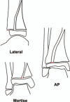
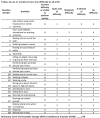
We assessed factors associated with premature physeal closure (PPC) and outcomes after closed reduction of Salter-Harris type II (SH-II) fractures of the distal tibia. We reviewed patients with SH-II fractures of the distal tibia treated at our center from 2010 to 2015 with closed reduction and a non-weightbearing long-leg cast. Patients were categorized by immediate postreduction displacement: minimal, <2 mm; moderate, 2 to 4 mm; or severe, >4 mm. Demographic data, radiographic data, and Lower Extremity Functional Scale (LEFS) scores were recorded.Fifty-nine patients (27 girls, 31 right ankles, 26 concomitant fibula fractures) were included, with a mean (±SD) age at injury of 12.0 ± 2.2 years. Mean maximum fracture displacements were 6.6 ± 6.5 mm initially, 2.7 ± 2.0 mm postreduction, and 0.4 ± 0.7 mm at final follow-up. After reduction, displacement was minimal in 23 patients, moderate in 21, and severe in 15. Fourteen patients developed PPC, with no significant differences between postreduction displacement groups. Patients with high-grade injury mechanisms and/or initial displacement ≥4 mm had 12-fold and 14-fold greater odds, respectively, of PPC. Eighteen patients responded to the LEFS survey (mean 4.0 ± 2.1 years after injury). LEFS scores did not differ significantly between postreduction displacement groups (P = .61).The PPC rate in this series of SH-II distal tibia fractures was 24% and did not differ by postreduction displacement. Initial fracture displacement and high-grade mechanisms of injury were associated with PPC. LEFS scores did not differ significantly by postreduction displacement.Level of Evidence: Level IV, case series.
Conflict of interest statement
The authors have no conflicts of interest to disclose.
Figures


References
-
- Peterson CA, Peterson HA. Analysis of the incidence of injuries to the epiphyseal growth plate. J Trauma 1972;12:275–81. - PubMed
-
- Rosenbaum AJ, DiPreta JA, Uhl RL. Review of distal tibial epiphyseal transitional fractures. Orthopedics 2012;35:1046–9. - PubMed
-
- Spiegel PG, Cooperman DR, Laros GS. Epiphyseal fractures of the distal ends of the tibia and fibula. A retrospective study of two hundred and thirty-seven cases in children. J Bone Joint Surg Am 1978;60:1046–50. - PubMed
-
- Russo F, Moor MA, Mubarak SJ, et al. Salter-Harris II fractures of the distal tibia: does surgical management reduce the risk of premature physeal closure? J Pediatr Orthop 2013;33:524–9. - PubMed
-
- Karrholm J, Hansson LI, Svensson K. Prediction of growth pattern after ankle fractures in children. J Pediatr Orthop 1983;3:319–25. - PubMed
MeSH terms
LinkOut - more resources
Full Text Sources
Miscellaneous

