Review
. 2020 May;96(3):478-499.
doi: 10.1111/php.13245. Epub 2020 May 4.

Deciphering UV-induced DNA Damage Responses to Prevent and Treat Skin Cancer

Affiliations
Review

Deciphering UV-induced DNA Damage Responses to Prevent and Treat Skin Cancer

Jihoon W Lee et al. Photochem Photobiol. 2020 May.

Abstract

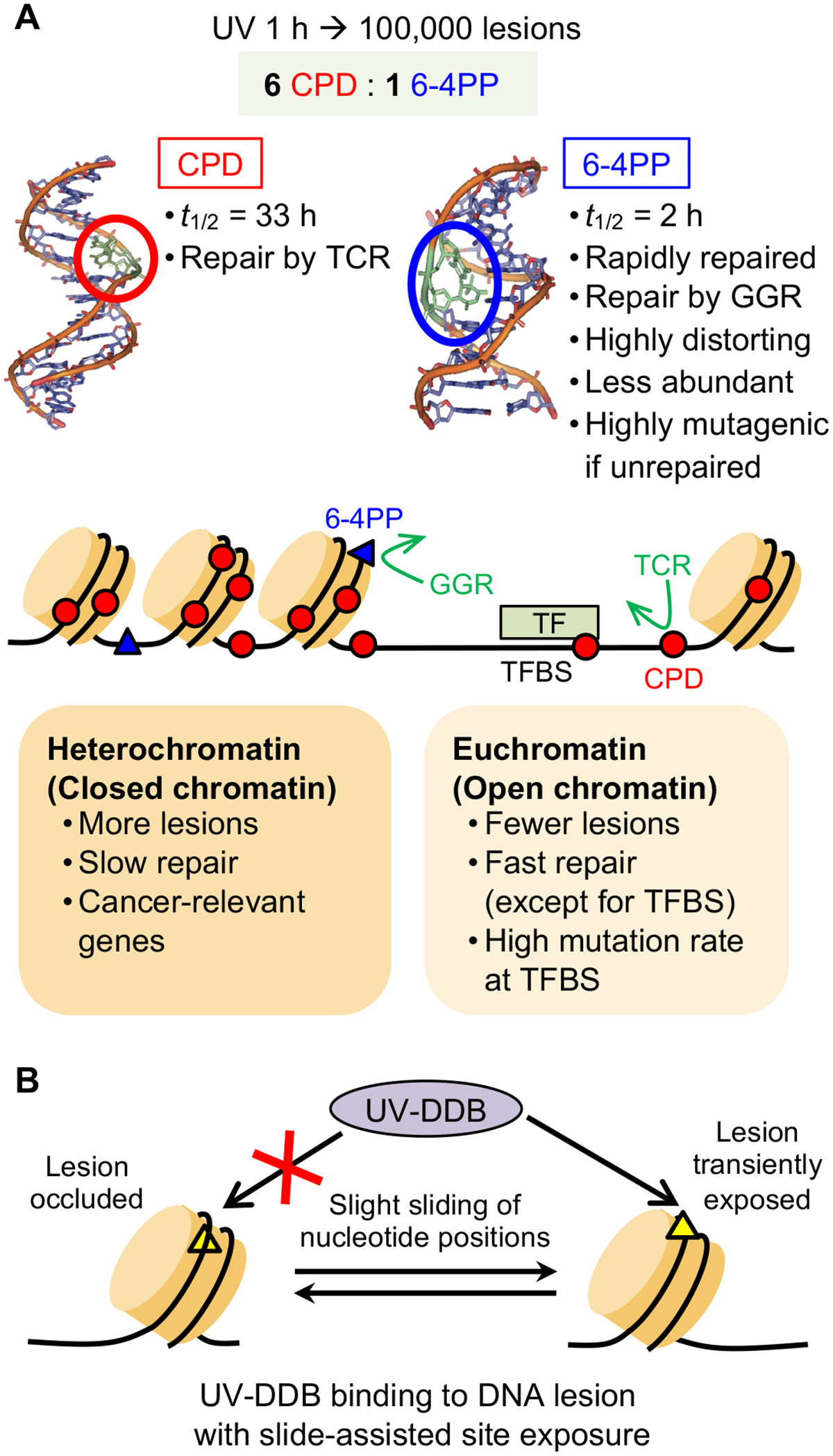
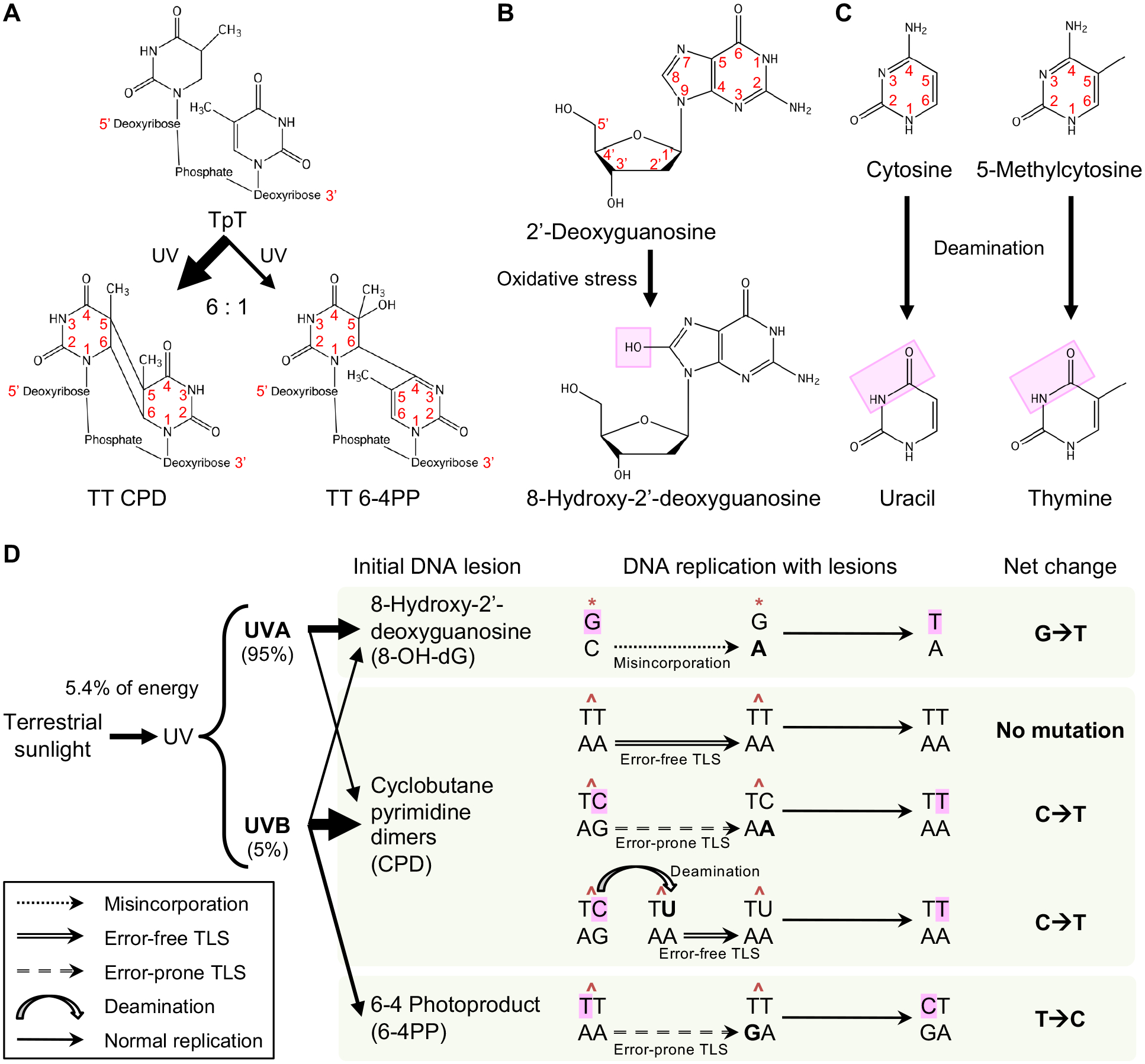
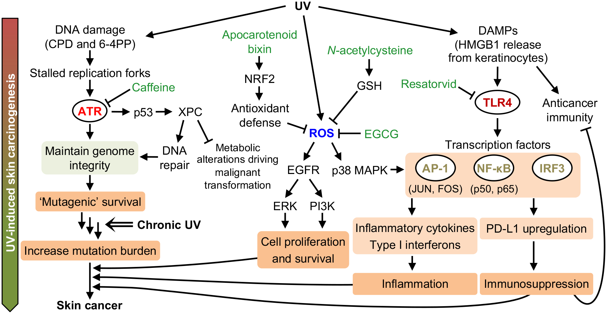
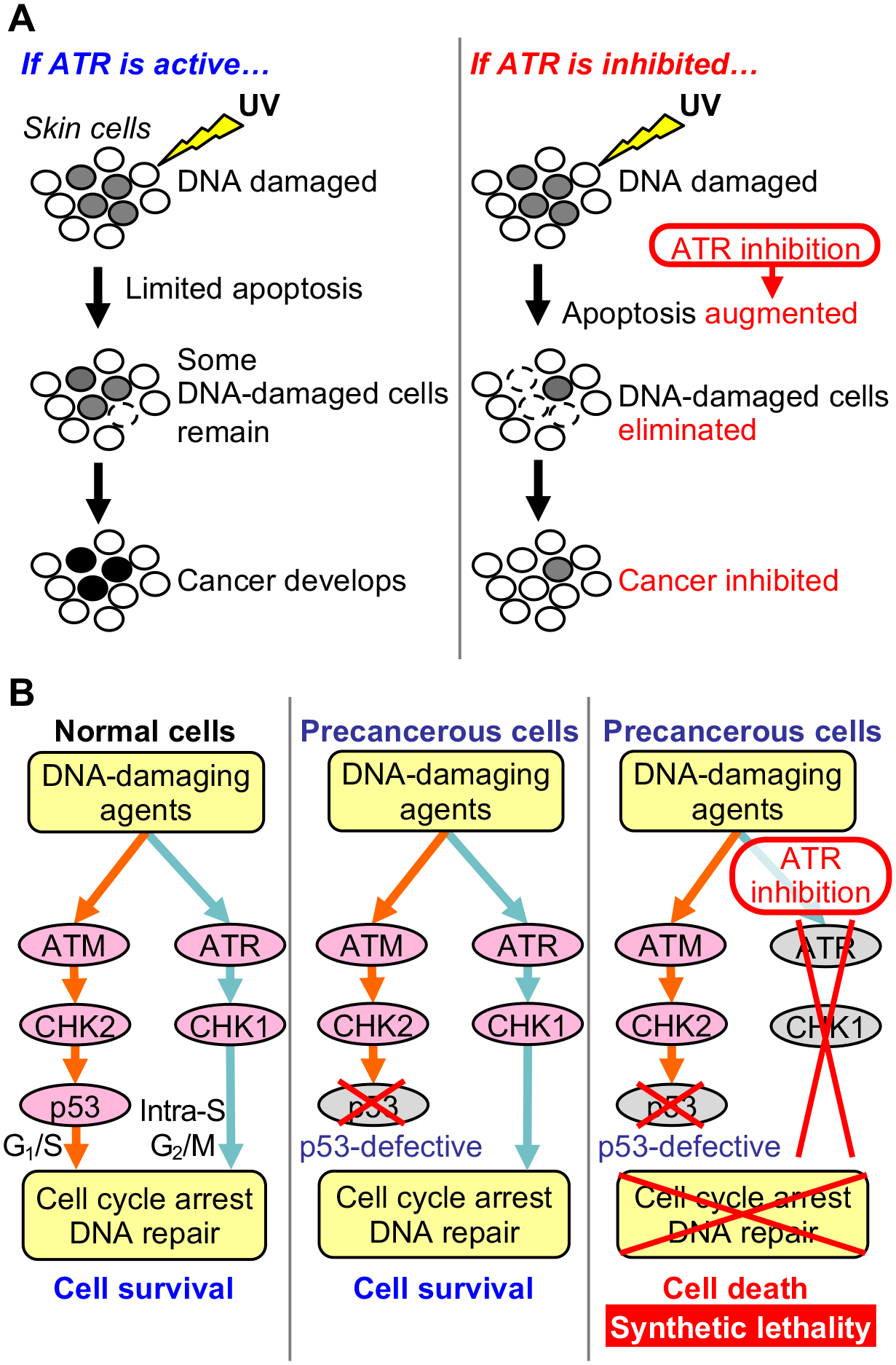
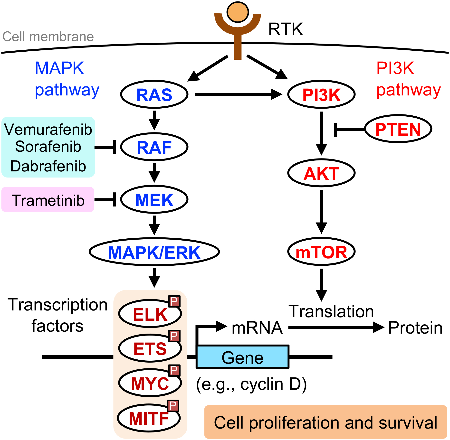
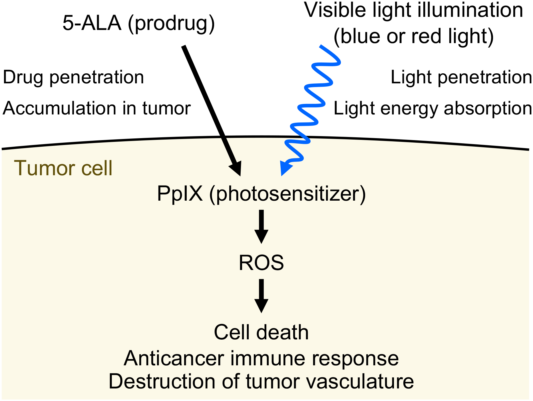
Ultraviolet (UV) radiation is among the most prevalent environmental factors that influence human health and disease. Even 1 h of UV irradiation extensively damages the genome. To cope with resulting deleterious DNA lesions, cells activate a multitude of DNA damage response pathways, including DNA repair. Strikingly, UV-induced DNA damage formation and repair are affected by chromatin state. When cells enter S phase with these lesions, a distinct mutation signature is created via error-prone translesion synthesis. Chronic UV exposure leads to high mutation burden in skin and consequently the development of skin cancer, the most common cancer in the United States. Intriguingly, UV-induced oxidative stress has opposing effects on carcinogenesis. Elucidating the molecular mechanisms of UV-induced DNA damage responses will be useful for preventing and treating skin cancer with greater precision. Excitingly, recent studies have uncovered substantial depth of novel findings regarding the molecular and cellular consequences of UV irradiation. In this review, we will discuss updated mechanisms of UV-induced DNA damage responses including the ATR pathway, which maintains genome integrity following UV irradiation. We will also present current strategies for preventing and treating nonmelanoma skin cancer, including ATR pathway inhibition for prevention and photodynamic therapy for treatment.


Conflict of interest statement

Competing Interests: The authors declare no competing interests.

Figures

Figure 1.
Figure 1.
Time course of UV-induced responses in the skin and skin cancer evolution. UV irradiation instantaneously generates DNA lesions that are potentially mutagenic and carcinogenic. To survive UV damage, cells cope with these deleterious lesions by activating a variety of signaling pathways including ATR (DNA damage response). UV also generates reactive oxygen species (ROS) that promote inflammation and carcinogenesis. Translesion synthesis enables DNA synthesis in the presence of UV-induced DNA lesions but may incorporate mutations. In the days and weeks following UV exposure, the skin exhibits tissue-level responses such as erythema, tanning, angiogenesis, epidermal hyperplasia, and immunosuppression. Chronic UV exposure leads to accumulation of genetic and epigenetic alterations. As a result, aberrant cell signaling drives transformation of normal skin cells to premalignant lesions (e.g., actinic keratosis (AK) or benign melanocytic nevi). With further increase in the burden of genetic and epigenetic alterations, cells may evolve into invasive and metastatic skin cancer. Chronic UV irradiation also promotes premature skin aging (photoaging), characterized by wrinkle formation and reduced elasticity.
Figure 2.
Figure 2.
UV-induced DNA lesion formation affected by nucleotide positioning in nucleosome. Within an individual nucleosome, due to the rotation of nucleotides within the DNA double helix, nucleotides are periodically positioned close to (‘inner’) or far from (‘outer’) the histone core every ~10 bp. Compared to the ‘outer’ positions, nucleotides at the ‘inner’ positions have constrained flexibility. Thus, UV induces dimer formation more frequently at ‘outer’ dipyrimidines where DNA bending is more flexible.
Figure 3.
Figure 3.
UV-induced DNA damage responses to maintain genome integrity. (A) ATR activation by replication blockage. During DNA replication, DNA polymerase stalls at UV-induced DNA lesions while MCM helicase continues to unwind duplex DNA. This uncoupling of helicase and polymerase activities generates a long stretch of single-stranded DNA (ssDNA) that is rapidly coated with RPA. RPA-coated ssDNA recruits the ATR-ATRIP complex, TopBP1, RHINO, and ETAA1, resulting in ATR activation. ATR phosphorylates many downstream targets to induce cell cycle arrest, inhibit DNA replication origin firing, and prevent replication fork collapse. (B) ATM activation by transcription blockage. In noncycling cells, UV-induced DNA lesions stall transcription and displace spliceosomes. This leads to hybridization of pre-mRNA with the template DNA strand, leaving the nontemplate strand as ssDNA and forming an R-loop structure that recruits ATM. Subsequently, ATM induces alternative splicing of pre-mRNA, leading to altered gene expression in UV-irradiated cells. (C) Nucleotide excision repair (NER) for UV-induced DNA lesions. NER consists of global genome repair (GGR), which recognizes lesions throughout the genome independently of transcription or replication, and transcription-coupled repair (TCR), which recognizes DNA lesions on transcribed strands of active genes during transcription. In both GGR and TCR, the damaged DNA strand is cleaved at both sides of the DNA lesion, generating a lesion-containing product (~30 nt). The resulting ssDNA gap is filled by DNA polymerases δ/ε. (D) EXO1-mediated ATR activation and response to excessive lesions. In noncycling cells, EXO1 competes with NER and extends the NER-generated ssDNA gap, making it long enough to activate the DNA damage checkpoint. Compared to low doses, high doses of UV irradiation generate lesions more frequently, and thus there is a greater chance that two DNA lesions are closely positioned on opposing strands (closely opposing lesions, COLs). One lesion is removed by NER, but the other lesion remains in the resulting ssDNA gap. NER gap filling by DNA polymerases δ/ε stalls at this lesion, and EXO1 extends the ssDNA gap. Translesion synthesis (TLS) polymerases fill the lesion-containing gap and displace EXO1. If TLS polymerases are deficient, EXO1 will continue to extend the ssDNA gap, leading to double-strand breaks and cell death. (E) Translesion synthesis (TLS) as DNA damage tolerance. When cells enter S phase with UV-induced DNA lesions, replicative DNA polymerases stall at the lesion. To resume DNA replication, repriming occurs downstream of the lesion. The resulting ssDNA gap will be filled by TLS polymerases that can synthesize DNA across the lesion but may incorporate mutations. TLS occurs in an error-free or error-prone manner.
Figure 4.
Figure 4.
Cell cycle checkpoints elicited by UV-induced ATR activation. G1-S checkpoint: Following UV-induced DNA damage in G1 phase, activated ATR phosphorylates p53 at Ser15 and CHK1 at Ser345. Phospho-CHK1 inactivates CDC25A, preventing dephosphorylation of CDK2 and inducing G1 arrest. The p53-p21 pathway also inhibits CDK2. Intra-S checkpoint: UV activates the ATR-CHK1 pathway in S phase, decreasing the function of CDK. DNA replication origin firing requires phosphorylation of Treslin by CDK, and thus it is plausible that replication stress prevents origin firing via inhibiting Treslin phosphorylation. S-G2 checkpoint: ATR senses ongoing DNA replication during unperturbed S phase, inhibiting CDK1 activity and preventing cell cycle progression to G2 phase. When DNA replication is completed, ATR activity is diminished, allowing CDK1 to phosphorylate FOXM1 for S-G2 transition. G2-M checkpoint: UV-induced ATR-CHK1 activation inhibits CDC25C and CDK1 activities, preventing G2-M transition. WEE1 phosphorylates and inhibits CDK1, also inducing G2 arrest.
Figure 5.
Figure 5.
Repair of UV-induced DNA lesions depends on chromatin state and sliding of nucleotide positions. (A) UV irradiation forms dimers at dipyrimidine sites on the same strand, distorting the DNA double helix. The two major types of DNA lesions are cyclobutane pyrimidine dimers (CPDs) and 6–4 photoproducts (6–4PPs), which differ in terms of their abundance, degree of DNA distortion, and the primary mode of repair (global genome repair (GGR) or transcription-coupled repair (TCR)). Chromatin state influences formation and repair of UV-induced DNA lesion; lesions form more frequently and are repaired more slowly in heterochromatin than in euchromatin. Transcription factor binding sites (TFBS) have variable frequency of lesion formation and slow lesion repair that leads to high mutation rate. The structures of DNA double helix with CPD and 6–4PP lesions are adapted with permission from Rastogi, R. P. et al. J Nucleic Acids (2010) 2010:592980. (B) UV-induced DNA lesions that are occluded within a nucleosome undergo ‘slide-assisted site exposure’: slight sliding of nucleotide positions relative to the histone core, without affecting the overall nucleosome architecture. This process transiently exposes occluded lesions to UV-damaged DNA-binding protein (UV-DDB), which recognizes UV damage and initiates global genome repair (GGR).
Figure 6.
Figure 6.
Chemical structures of UV-induced DNA lesions and mechanisms of UV-induced mutagenesis. (A) Cyclobutane pyrimidine dimer (CPD) and 6–4 photoproduct (6–4PP) lesions are UV-induced dimers formed at dipyrimidine sites, e.g., two adjacent thymines (TpT). CPDs are generated by UV from simulated sunlight 6 times more frequently than 6–4PPs. (B) Oxidative stress can be induced by UVA rather than UVB and damages 2’-deoxyguanosine, converting it into the DNA lesion 8-hydroxy-2’-deoxyguanosine (8-OH-dG). (C) Cytosine and 5-methylcytosine (5-mC) can be deaminated into uracil and thymine, respectively. Deamination occurs more frequently when cytosine or 5-mC is part of a CPD on a transcribed strand. (D) UVA and UVB compose 95% and 5% of UV radiation from terrestrial sunlight, respectively. UVA and UVB irradiation generate 8-OH-dG, CPD, and 6–4PP DNA lesions. Deamination may occur at CPDs. During the first round of DNA replication after lesion formation, correct or incorrect nucleotides are incorporated opposite of the lesion via different mechanisms including misincorporation and error-free and error-prone translesion synthesis (TLS). During the second round of DNA replication, complementary nucleotides are incorporated opposite of the correct or incorrect nucleotides that were from the first round. This results in mutation fixation. Due to the abundance and slow repair of CPDs, the resulting C>T mutations are the most prevalent UV-induced mutations and are known as UV signature mutations. Thickness of each arrow leading from UVA and UVB to DNA lesion types in the panel indicates the relative contributions of UVA and UVB to formation of each lesion type.
Figure 7.
Figure 7.
Targeting UV-induced signaling pathways to suppress skin carcinogenesis. UV-induced DNA lesions stall DNA replication forks and activate the ATR pathway. The ATR pathway maintains genome integrity, allowing cells to survive UV-induced DNA damage but possibly with unrepaired lesions that become mutations (‘mutagenic survival’). Thus, chronic UV exposure will increase mutation burden, accumulating cancer driver mutations that may promote skin cancer development. UV-induced p53 upregulates XPC, which facilitates DNA repair and prevents metabolic alterations that drive cancer. UV-induced reactive oxygen species (ROS) activate the EGFR and p38 MAPK pathways, contributing to skin cancer development. HMGB1 released from UV-damaged keratinocytes activates the TLR4 pathway, which can drive skin cancer via inflammation and immunosuppression. Inhibiting ATR (via caffeine), ROS (via the tea polyphenol EGCG), or TLR4 (via resatorvid) has been demonstrated to prevent UV-induced skin carcinogenesis in vivo.
Figure 8.
Figure 8.
Targeting the ATR pathway to prevent cancer development. (A) ATR inhibition prevents UV-induced skin cancer development. After UV exposure, ATR induces cell cycle arrest that allows time for DNA repair, resulting in limited apoptosis. However, when DNA replication is ongoing before completion of DNA repair, mutations may be incorporated via DNA damage tolerance mechanisms (‘mutagenic survival’), leading to skin cancer development. In contrast, inhibition of the ATR pathway that is essential for surviving UV damage leads to augmented apoptosis. This contributes to elimination of DNA-damaged cells that are at increased risk of malignant transformation, thereby inhibiting skin cancer development. (B) ATR inhibition sensitizes p53-defective cells to DNA-damaging agents. Normal cells have two major pathways that respond to DNA damage: the ATM-CHK2-p53 and ATR-CHK1 pathways, which induce cell cycle arrest and DNA repair. However, precancerous cells are often defective in p53 and thus rely on the ATR-CHK1 pathway to survive DNA damage. When p53-defective precancerous cells are treated with an ATR inhibitor in the presence of a DNA-damaging agent (e.g., UV or chemotherapeutic drug), these cells will have no intact DNA damage response pathway to survive the DNA damage (synthetic lethality of defective p53 and ATR inhibition). As a result, precancerous cells will undergo apoptosis, and cancer development will be suppressed. This synthetic lethality provides the molecular basis for the use of ATR inhibitors to prevent cancer development from p53-defective cells.
Figure 9.
Figure 9.
Targeting oncogenic MAPK and PI3K pathways in skin cancer. Receptor tyrosine kinases (RTKs) activate the mitogen-activated protein kinase/extracellular signal-regulated kinase (MAPK/ERK) pathway and the phosphoinositide 3-kinase (PI3K) pathway, which are frequently aberrant in skin cancer. MAPK/ERK phosphorylates transcription factors and upregulates expression of genes important for cell proliferation and survival. The mammalian target of rapamycin (mTOR) upregulates mRNA translation of these genes. Aberrant activation of these pathways (e.g., activating BRAFV600E mutation) leads to increased proliferation and survival. RAF inhibitors, such as vemurafenib, sorafenib, and dabrafenib, and the MEK inhibitor trametinib are currently used in melanoma treatment. However, RAF inhibitor monotherapy for melanoma can accelerate the progression of preexisting RAS-mutant skin lesions into cutaneous squamous cell carcinoma (cSCC). Combination therapy with dabrafenib and trametinib, each targeting a different kinase within the MAPK pathway, reduces the incidence of cSCC in melanoma patients treated with a RAF inhibitor.
Figure 10.
Figure 10.
Mechanism of photodynamic therapy (PDT) for cancer. The prodrug 5-aminolevulinic acid (5-ALA) is preferentially distributed to tumor tissue in a patient. Tumor cells metabolize 5-ALA into the photosensitizer protoporphyrin IX (PpIX). Subsequently, target cells are exposed to visible light (either 400 nm or 635 nm) that is absorbed by PpIX. Excited PpIX generates reactive oxygen species (ROS) that lead to tumor cell death, anticancer immune response, and destruction of tumor vasculature. Visible light illumination and the prodrug 5-ALA have limited penetration into deeper tumor tissue, reducing the efficacy of PDT for larger tumors.

Similar articles

Cited by

References

    1. Kwa RE, Campana K and Moy RL (1992) Biology of cutaneous squamous cell carcinoma. J Am Acad Dermatol 26, 1–26. - PubMed
    1. Albert MR and Ostheimer KG (2002) The evolution of current medical and popular attitudes toward ultraviolet light exposure: part 1. J Am Acad Dermatol 47, 930–937. - PubMed
    1. Armstrong BK, Kricker A and English DR (1997) Sun exposure and skin cancer. Australas J Dermatol 38 Suppl 1, S1–6. - PubMed
    1. Wacker M and Holick MF (2013) Sunlight and Vitamin D: A global perspective for health. Dermatoendocrinol 5, 51–108. - PMC - PubMed
    1. Clydesdale GJ, Dandie GW and Muller HK (2001) Ultraviolet light induced injury: immunological and inflammatory effects. Immunol Cell Biol 79, 547–568. - PubMed

Publication types