. 2020 Mar 16;33(3):751-763.
doi: 10.1021/acs.chemrestox.9b00363. Epub 2020 Mar 2.

Stem Cell-Derived Endothelial Cell Model that Responds to Tobacco Smoke Like Primary Endothelial Cells

Affiliations

Stem Cell-Derived Endothelial Cell Model that Responds to Tobacco Smoke Like Primary Endothelial Cells

Pei-Hsuan Chu et al. Chem Res Toxicol. .

Abstract

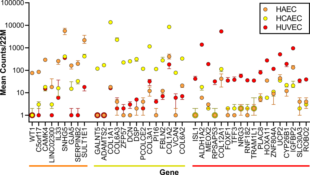
To clarify how smoking leads to heart attack and stroke, we developed an endothelial cell model (iECs) generated from human induced Pluripotent Stem Cells (iPSC) and evaluated its responses to tobacco smoke. These iECs exhibited a uniform endothelial morphology, and expressed markers PECAM1/CD31, VWF/ von Willebrand Factor, and CDH5/VE-Cadherin. The iECs also exhibited tube formation and acetyl-LDL uptake comparable to primary endothelial cells (EC). RNA sequencing (RNA-Seq) revealed a robust correlation coefficient between iECs and EC (R = 0.76), whereas gene responses to smoke were qualitatively nearly identical between iECs and primary ECs (R = 0.86). Further analysis of transcriptional responses implicated 18 transcription factors in regulating responses to smoke treatment, and identified gene sets regulated by each transcription factor, including pathways for oxidative stress, DNA damage/repair, ER stress, apoptosis, and cell cycle arrest. Assays for 42 cytokines in HUVEC cells and iECs identified 23 cytokines that responded dynamically to cigarette smoke. These cytokines and cellular stress response pathways describe endothelial responses for lymphocyte attachment, activation of coagulation and complement, lymphocyte growth factors, and inflammation and fibrosis; EC-initiated events that collectively lead to atherosclerosis. Thus, these studies validate the iEC model and identify transcriptional response networks by which ECs respond to tobacco smoke. Our results systematically trace how ECs use these response networks to regulate genes and pathways, and finally cytokine signals to other cells, to initiate the diverse processes that lead to atherosclerosis and cardiovascular disease.


Figures

Figure 1.
Figure 1.. Generation of endothelial cells from human iPS cells in fully defined conditions.
(A) iPSCs (1 × 10⁵/well in 6-well microplate) were seeded onto Matrigel-coated plates with E8 Medium at day 0, and then the medium was changed to Modified Dulbecco’s Media (MDM) on the next day (day 1). Scale bars show 100 microns. After 7 days induction, PECAM1-positive cells were evaluated for endothelial marker proteins and phenotypes. (B) The “cobblestone” morphology of the iECs was maintained after multiple passages.
Figure 2.
Figure 2.. Evaluation of iPSC- derived endothelial cells (iECs) morphology and phenotypes.
(A) iECs derived from iPSC through mesoderm precursors showed typical endothelial cell morphology, and expressed characteristic EC markers (CD31/PECAM1, vWF/von Willebrand Factor, and CDH5, red) defined by immunofluorescent staining and FACS. Immunostaining used phycoerythrin or allophycocyanin (PE or APC). Nuclei were visualized with DAPI (Blue). Image bar: 100 μm. (B) In vitro angiogenesis, or tube formation functional assay for iPSC-derived iECs. iECs cultured on Matrigel formed vascular tube-like structures. Image bar: 200 μm. The total number of junctions and branch lengths of networks is shown (N=5). (C) The capacities of iEC or HUVEC for acetyl-LDL (Dil-Ac-LDL) uptake was determined by quantifying fluorescence in images captured by a microscope (image bar: 100 μm) and FACS (plots; N=5).
Figure 3.
Figure 3.. Genes expressed selectively in EC from human aorta, coronary artery, or umbilicus.
RNAseq data are shown for genes expressed at ten-fold higher counts in HAEC (orange) or HUVEC (red), or twenty-fold higher in HCAEC (yellow) relative to the average of the other two EC beds. For each gene the median number of transcripts per 22 million is plotted +/− the standard error for each EC bed. Zero transcript counts were plotted as 1 transcript to fit the logarithmic scale.
Figure 4.
Figure 4.. Gene expression comparison of iECs to primary endothelial cells.
(A) PCA relating gene expression profiles of vehicle-treated fibroblasts, blue squares; iPSCs, black triangles; HAEC, orange circles; HCAEC, yellow circles; HUVEC, red circles; and two iEC lines, green diamonds. Three biological replicates represent each cell line. (B) Scatter plot comparing baseline gene expression profiles of the mean expression level for the three primary cell beds and iEC lines. Squares represent genes that deviate by 3 standard deviations from the least squares regression line (gray).
Figure 5.
Figure 5.. Gene expression responses to tobacco smoke, comparing iECs to primary endothelial cells.
(A) PCA relating gene expression responses of the EC cell lines depicted in Figure 3 to tobacco smoke for six hours. Circles represent vehicle-treated cells, and triangles represent smoke-treated cells. (B) Heat maps comparing 8,840 genes that responded significantly to tobacco smoke in any of the five cell lines (left side). The right-side heat map shows only the 2,285 genes that responded significantly to smoke in both iECs or in all three primary EC types. Genes that increased or decreased in response to smoke are shown in red or green, respectively, with color saturation at 4-fold. Genes that did not change in response to smoke are shown in black, and genes with <25 normalized counts per sample are shown in grey. (C) Scatter plot showing gene expression responses to smoke, comparing iECs to mean values for the three primary EC types. Axes show Log2 fold-change values for smoke/vehicle-treatments. This scatterplot shows a spot for each of the 10,804 genes that yielded at least 25 counts from each cell type for either vehicle or smoke treatment. Black spots represent genes that responded significantly to smoke in three or more cell lines. The coefficient of correlation for the regression line R = 0.86.
Figure 6.
Figure 6.. Eighteen TFs implicated in responses to smoke by primary ECs and iECs.
The heat map shows TF-regulated gene sets from Enrichr that overlapped with the 762 smoke responsive genes that responded to all three primary ECs or both iEC lines. These eighteen TF-associated gene sets showed significant overlap in two or more experimental sources in Enrichr. Light blue lines mark genes with direct cis-regulation of the gene by the TF (response element, ChIP-seq, or ChIP-chip). Black lines mark genes that were empirically observed to be regulated by the indicated TF, possibly indirectly in trans, in two datasets. Dark blue lines indicate both direct and empirical evidence. A dendrogram indicates intersecting gene sets among TFs. Among the TFs, the NFE2L2 gene encodes NRF2, ESR1 encodes the Estrogen Receptor α, and RELA and RELB encode NFκB family TFs.
Figure 7.
Figure 7.. Cytokines secreted by HUVECs and iECs in response to tobacco smoke.
Bar graphs show 29 cytokines in control conditions (white bars) and their responses to smoke (black bars) in HUVECs or iECs. Cytokines are grouped into three charts according to protein concentrations in the medium, below 50 pg/mL, 50–600 pg/mL, and above 600 pg/mL. An asterisk indicates a significant response to smoke p < 0.05.

References

    1. Hoffmann D, Djordjevic MV, and Hoffmann I. (1997) The changing cigarette. Prev Med 26, 427–434. - PubMed
    1. Alberg AJ, Shopland DR, and Cummings KM (2014) The 2014 Surgeon General’s report: commemorating the 50th Anniversary of the 1964 Report of the Advisory Committee to the US Surgeon General and updating the evidence on the health consequences of cigarette smoking. Am J Epidemiol 179, 403–412. - PMC - PubMed
    1. Backinger CL, Meissner HI, and Ashley DL (2016) The FDA “Deeming Rule” and Tobacco Regulatory Research. Tob Regul Sci 2, 290–293. - PMC - PubMed
    1. Messner B, and Bernhard D. (2014) Smoking and cardiovascular disease: mechanisms of endothelial dysfunction and early atherogenesis. Arterioscler Thromb Vasc Biol 34, 509–515. - PubMed
    1. Leone A. (2015) Toxics of Tobacco Smoke and Cardiovascular System: From Functional to Cellular Damage. Curr Pharm Des 21, 4370–4379. - PubMed

Publication types

MeSH terms