. 2020 Mar 2;15(3):e0229240.
doi: 10.1371/journal.pone.0229240. eCollection 2020.

Apolipoprotein E regulates the maturation of injury-induced adult-born hippocampal neurons following traumatic brain injury

Affiliations

Apolipoprotein E regulates the maturation of injury-induced adult-born hippocampal neurons following traumatic brain injury

Yacine Tensaouti et al. PLoS One. .

Abstract

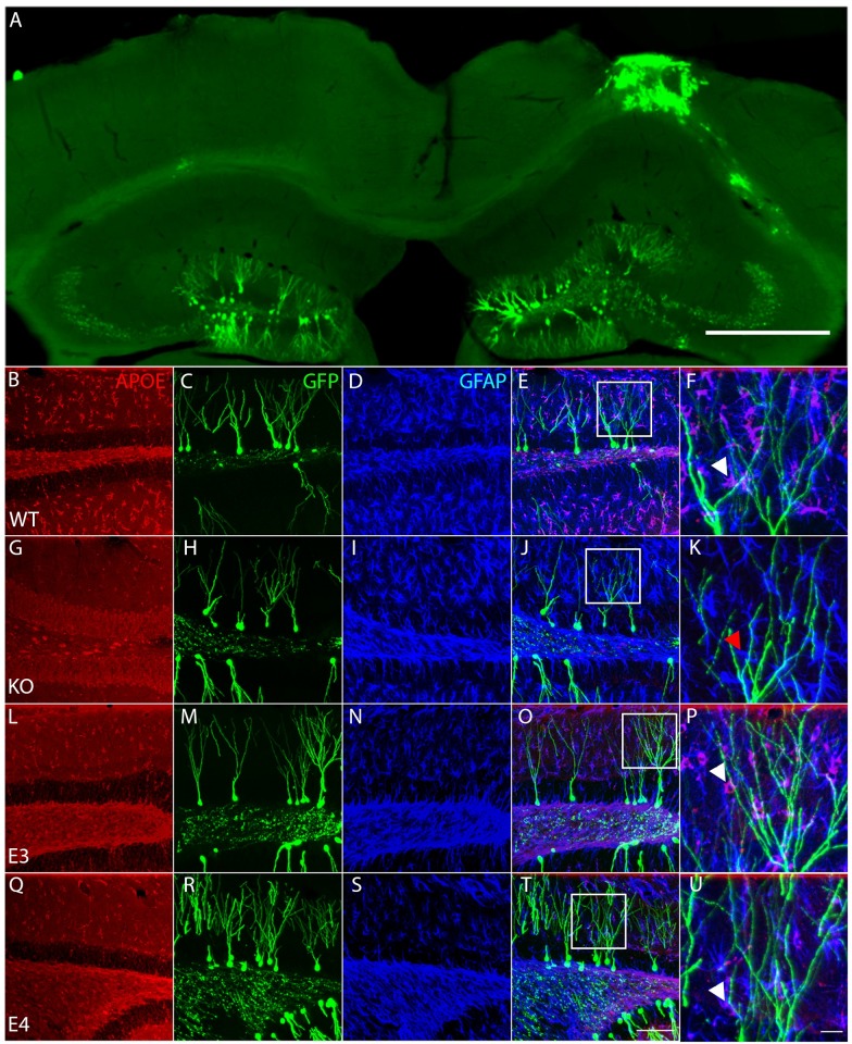
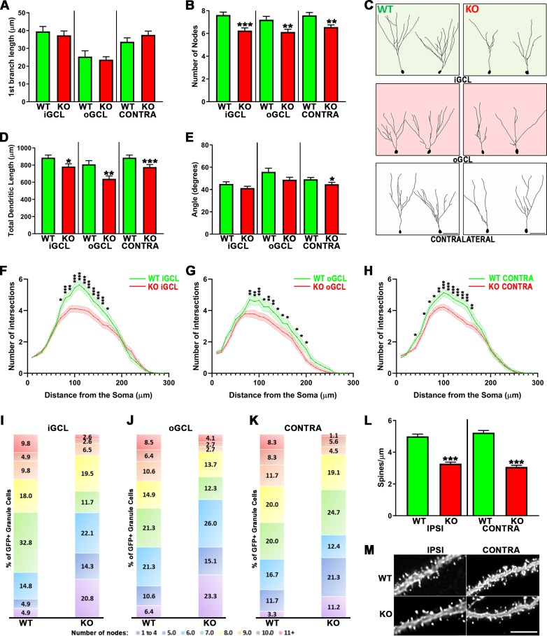
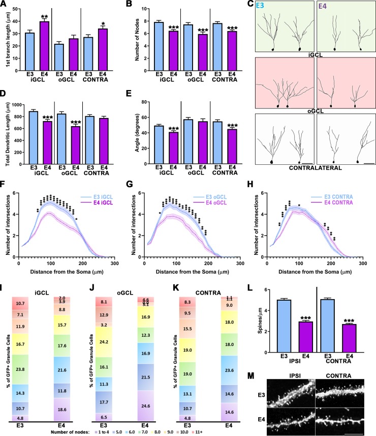
Various brain injuries lead to the activation of adult neural stem/progenitor cells in the mammalian hippocampus. Subsequent injury-induced neurogenesis appears to be essential for at least some aspects of the innate recovery in cognitive function observed following traumatic brain injury (TBI). It has previously been established that Apolipoprotein E (ApoE) plays a regulatory role in adult hippocampal neurogenesis, which is of particular interest as the presence of the human ApoE isoform ApoE4 leads to significant risk for the development of late-onset Alzheimer's disease, where impaired neurogenesis has been linked with disease progression. Moreover, genetically modified mice lacking ApoE or expressing the ApoE4 human isoform have been shown to impair adult hippocampal neurogenesis under normal conditions. Here, we investigate how controlled cortical impact (CCI) injury affects dentate gyrus development using hippocampal stereotactic injections of GFP-expressing retroviruses in wild-type (WT), ApoE-deficient and humanized (ApoE3 and ApoE4) mice. Infected adult-born hippocampal neurons were morphologically analyzed once fully mature, revealing significant attenuation of dendritic complexity and spine density in mice lacking ApoE or expressing the human ApoE4 allele, which may help inform how ApoE influences neurological diseases where neurogenesis is defective.


Conflict of interest statement

The authors have declared that no competing interests exist.

Figures

Fig 1
Fig 1. ApoE-expressing astrocytes physically interact with injury-induced GFP-expressing dendrites.
(A) Representative section of the contralateral and ipsilateral cortex and hippocampus 4 weeks after Controlled Cortical Impact and stereotaxic injection of GFP-expressing retroviruses in the dentate gyrus of a wildtype mouse reveals the typical morphology of the injured hippocampus along with efficient long-term labelling of neural progenitor cells. Scale bar 500μm. (B-U) Representative pictures of ApoE (first column, antigen is recombinant human ApoE and can react with human e2, e3 and e4, as well as non-human primate and mouse ApoE), GFP (second column) and GFAP (third column) immunostaining in the dentate gyrus of WT (B-F), ApoE KO (G-K), ApoE3 (L-P) and ApoE4 mice (Q-U). (E, J, O, T) Merged pictures (scale bar 100μm), white squares indicate enlarged area shown in last column (F, K, P, U), white arrowheads designate ApoE-expressing GFAP-positive astrocytes wrapping around GFP-expressing dendrites, red arrowhead designate ApoE-deficient GFAP-positive astrocyte, scale bar 10μm.
Fig 2
Fig 2. ApoE deficiency leads to impaired dendritic development of injury-induced adult-born granule neurons.
(A) ApoE deficiency did not affect the distance to the first dendritic branching but did significantly affect dendritic complexity (B) in the iGCL (p < 0.001), oGCL (p < 0.01) and contralateral (p < 0.01) dentate gyrus when compared to WT. (C) Representative tracings of GFP-expressing cells found in the iGCL, oGCL and contralateral dentate gyrus from WT and ApoE-deficient injured dentate gyrus. (D) The cumulative dendritic length is significantly attenuated in mouse lacking ApoE when compared to matching Wild Type cells (iGCL: p < 0.05, oGCL: p < 0.01, Contra: p < 0.001). (E) Injury-induced cells found in the contralateral dentate gyrus in the absence of ApoE also showed decreased dendritic span when compared to WT (p < 0.05). Sholl analysis of dendritic arborizations of CCI-induced adult-born granule neurons exposed differences in the dendritic branching number observed in both proximal and distal regions when comparing WT with ApoE-deficient GFP-expressing cells found in the iGCL (F), in the oGCL (G) and in the contralateral dentate gyrus (H). (I-K) In each condition, all traced neurons have been categorized depending on their complexity (≤4…≥11 nodes), highlighting higher proportions of less complex neurons (<4 nodes) in ApoE-deficient, particularly in the oGCL. 4 mice/condition and at least 10 neurons/mouse were analyzed; WT iGCL: 61 cells; WT oGCL: 47 cells; WT Contra: 60 cells; ApoE KO iGCL: 77 cells; ApoE KO oGCL: 73 cells; ApoE KO Contra: 89 cells. Contra = Contralateral dentate gyrus; iGCL = Inner Granule Cell Layer, oGCL = Outer Granule Cell Layer, in the ipsilateral dentate gyrus; WT = Wild Type, KO = ApoE Knock-out. ***p < 0.001. (L) ApoE deficiency leads to significantly reduced spine density in injury-induced adult-born granule neurons when compared to WT cells from both the Ipsilateral and Contralateral sides (p < 0.001). High power representative pictures of dendritic fragments from mature adult-born granule neurons in wildtype and ApoE-deficient (M). 4 mice/condition; Number of dendritic fragments analyzed: WT Ipsilateral = 60, WT Contralateral = 62, ApoE KO Ipsilateral = 87, ApoE KO Contralateral = 53. Scale bar = 5μm.
Fig 3
Fig 3. Impaired dendritic development of adult-born granule neurons after CCI in ApoE4 mouse.
(A) GFP-cells found in the iGCL (p < 0.01) and contralateral dentate gyrus (p < 0.05) of ApoE4 mice have the first dendritic branching occurring further from their cell body when compared to ApoE3. (B) The number of dendritic intersections of adult-born granule cells located in the iGCL, oGCL and contralateral dentate gyrus of ApoE4 mice is significantly reduced when compared to ApoE3 (p < 0.001). (C) Representative tracings of GFP-expressing cells found in the iGCL, oGCL and contralateral dentate gyrus from ApoE3 and ApoE4 adult mouse injured dentate gyrus. (D) The cumulative length of ApoE4-expressing injury-induced neurons’ dendrites is significantly reduced in both the iGCL and oGCL (p < 0.001). (E) ApoE4 significantly impairs the dendritic span of adult-born granule cells from both the iGCL and the contralateral dentate gyrus (p < 0.001). Sholl analysis revealed significant differences in the number of dendritic intersections observed in both proximal and distal regions when comparing ApoE3 with ApoE4-expressing cells found in the iGCL (F), in the oGCL (G) and in the contralateral dentate gyrus (H). 4 mice/condition and at least 10 neurons/mouse were analyzed; ApoE3 iGCL: 81 cells; ApoE3 oGCL: 62 cells; ApoE3 Contra: 81 cells; ApoE4 iGCL: 102 cells; ApoE4 oGCL: 65 cells; ApoE4 Contra: 89 cells. Contra = Contralateral dentate gyrus; iGCL = Inner Granule Cell Layer, oGCL = Outer Granule Cell Layer, in the ipsilateral dentate gyrus. *p < 0.05, **p < 0.01, ***p < 0.001. (I-K) In each condition, all traced neurons have been categorized depending on their complexity (≤4…≥11 nodes), highlighting higher proportions of less complex neurons (<4 nodes) in ApoE4-expressing mice, particularly in the oGCL. 4 mice/condition and at least 10 neurons/mouse were analyzed; ApoE3 iGCL: 81 cells; ApoE3 oGCL: 62 cells; ApoE3 Contra: 84 cells; ApoE4 iGCL: 102 cells; ApoE4 oGCL: 65 cells; ApoE4 Contra: 82 cells. Contra = Contralateral dentate gyrus; iGCL = Inner Granule Cell Layer, oGCL = Outer Granule Cell Layer, in the ipsilateral dentate gyrus; E3 = ApoE3, E4 = ApoE4. ***p < 0.001. (L) ApoE4-expressing adult-born granule neurons show significantly attenuated spine density when compared to ApoE3 matching cells from both dentate gyrus (p < 0.001). High power representative pictures of dendritic fragments from mature adult-born granule neurons in ApoE3 and ApoE4 (M) ipsilateral and contralateral dentate gyrus. 4 mice/condition; Number of dendritic fragments analyzed: ApoE3 Ipsilateral = 99, ApoE3 Contralateral = 91, ApoE4 Ipsilateral = 71, ApoE4 Contralateral = 58. Scale bar = 5μm.

References

    1. Dewan MC, Rattani A, Gupta S, Baticulon RE, Hung YC, Punchak M, et al. Estimating the global incidence of traumatic brain injury. J Neurosurg. 2018:1–18. - PubMed
    1. Rubiano AM, Carney N, Chesnut R, Puyana JC. Global neurotrauma research challenges and opportunities. Nature. 2015;527(7578):S193–7. 10.1038/nature16035 - DOI - PubMed
    1. Blaiss CA, Yu TS, Zhang G, Chen J, Dimchev G, Parada LF, et al. Temporally specified genetic ablation of neurogenesis impairs cognitive recovery after traumatic brain injury. J Neurosci. 2011;31(13):4906–16. 10.1523/JNEUROSCI.5265-10.2011 - DOI - PMC - PubMed
    1. Gao X, Deng-Bryant Y, Cho W, Carrico KM, Hall ED, Chen J. Selective death of newborn neurons in hippocampal dentate gyrus following moderate experimental traumatic brain injury. J Neurosci Res. 2008;86(10):2258–70. 10.1002/jnr.21677 - DOI - PMC - PubMed
    1. Yu TS, Zhang G, Liebl DJ, Kernie SG. Traumatic brain injury-induced hippocampal neurogenesis requires activation of early nestin-expressing progenitors. J Neurosci. 2008;28(48):12901–12. 10.1523/JNEUROSCI.4629-08.2008 - DOI - PMC - PubMed

Publication types

MeSH terms