

Correlation between histogram-based DCE-MRI parameters and 18F-FDG PET values in oropharyngeal squamous cell carcinoma: Evaluation in primary tumors and metastatic nodes
- PMID: 32119697
- PMCID: PMC7051076
- DOI: 10.1371/journal.pone.0229611
Correlation between histogram-based DCE-MRI parameters and 18F-FDG PET values in oropharyngeal squamous cell carcinoma: Evaluation in primary tumors and metastatic nodes
Abstract
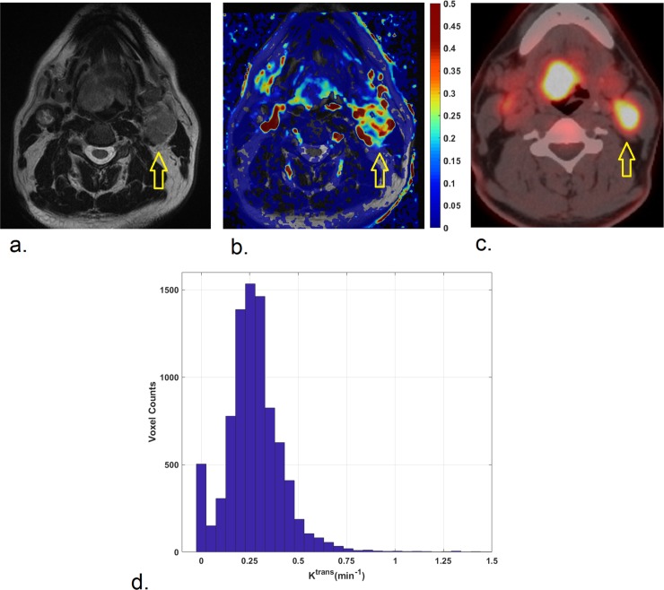
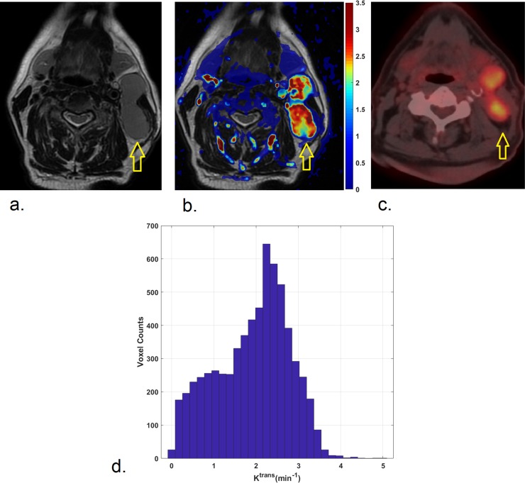
Objectives: To investigate the correlation between histogram-based Dynamic Contrast-Enhanced magnetic resonance imaging (DCE-MRI) parameters and positron emission tomography with 18F-fluorodeoxyglucose (18F-FDG-PET) values in oropharyngeal squamous cell carcinoma (OPSCC), both in primary tumors (PTs) and in metastatic lymph nodes (LNs).
Methods: 52 patients with a new pathologically-confirmed OPSCC were included in the present retrospective cohort study. Imaging including DCE-MRI and 18F-FDG PET/CT scans were acquired in all patients. Both PTs and the largest LN, if present, were volumetrically contoured. Quantitative parameters, including the transfer constants, Ktrans and Kep, and the volume of extravascular extracellular space, ve, were calculated from DCE-MRI. The percentiles (P), P10, P25, P50, P75, P90, and skewness, kurtosis and entropy were obtained from the histogram-based analysis of each perfusion parameter. Standardized uptake values (SUV), SUVmax, SUVpeak, SUVmean, metabolic tumor volume (MTV) and total lesion glycolysis (TLG) were calculated applying a SUV threshold of 40%. The correlations between all variables were investigated with the Spearman-rank correlation test. To exclude false positive results under multiple testing, the Benjamini-Hockberg procedure was applied.
Results: No significant correlations were found between any parameters in PTs, while significant associations emerged between Ktrans and 18F-FDG PET parameters in LNs.
Conclusions: Evident relationships emerged between DCE-MRI and 18F-FDG PET parameters in OPSCC LNs, while no association was found in PTs. The complex relationships between perfusion and metabolic biomarkers should be interpreted separately for primary tumors and lymph-nodes. A multiparametric approach to analyze PTs and LNs before treatment is advisable in head and neck squamous cell carcinoma (HNSCC).
Conflict of interest statement
The authors have declared that no competing interests exist.
Figures
References
Publication types
MeSH terms
Substances
LinkOut - more resources
Full Text Sources
Medical
Research Materials
