. 2020 Mar 2;15(3):e0229996.
doi: 10.1371/journal.pone.0229996. eCollection 2020.

Efficient immortalization of human dental pulp stem cells with expression of cell cycle regulators with the intact chromosomal condition

Affiliations

Efficient immortalization of human dental pulp stem cells with expression of cell cycle regulators with the intact chromosomal condition

Ai Orimoto et al. PLoS One. .

Abstract

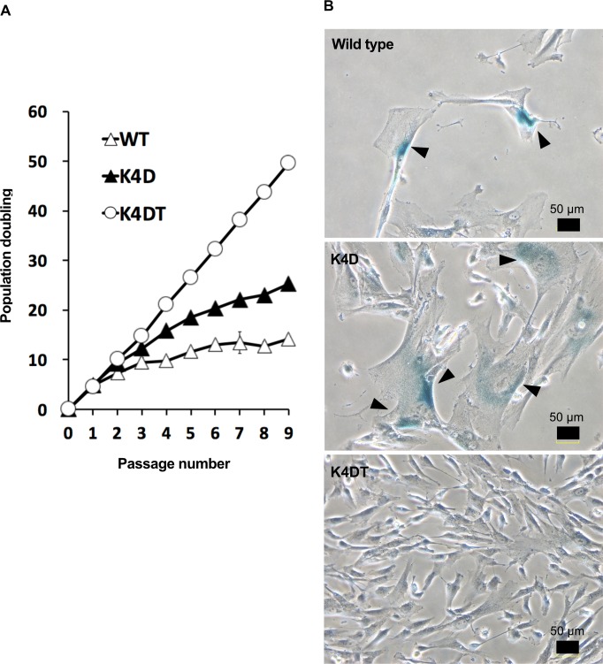
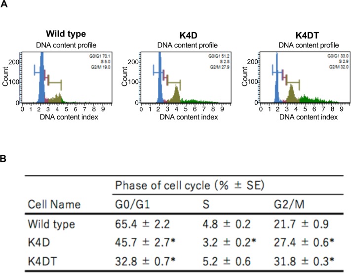
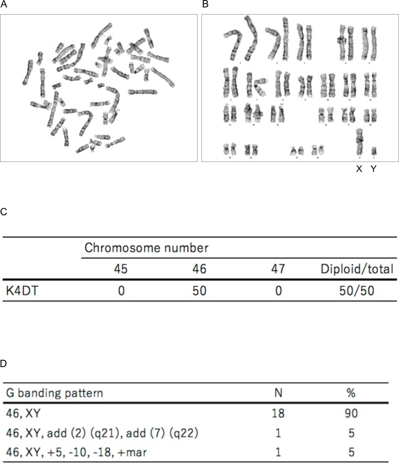
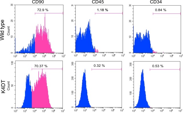
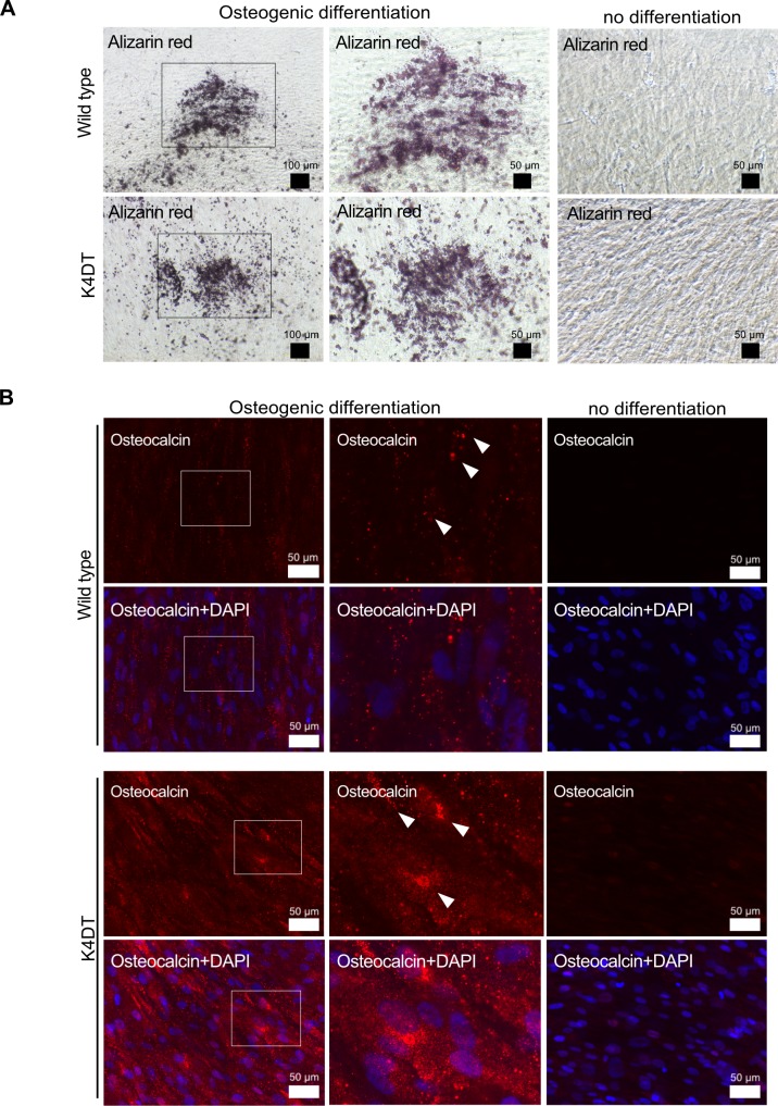
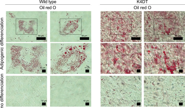
Clinical studies have recently demonstrated that autologous transplantation of mobilized dental pulp stem cells is a safe and efficacious potential therapy for pulp regeneration. However, some limitations need to be addressed, such as the high cost of the safety and quality control tests for isolated individual dental pulp cell products before transplantation. Therefore, more efficient in vitro culturing of human dental pulp stem cells might be useful for providing low cost and high reliability testing for pulp regeneration therapy. In this study, we established a novel immortalized dental pulp stem cell line by co-expressing a mutant cyclin-dependent kinase 4 (CDK4R24C), Cyclin D1, and telomerase reverse transcriptase (TERT). The established cell line maintained its original diploid chromosomes and stemness characteristics and exhibited an enhanced proliferation rate. In addition, we showed the immortalized human dental pulp stem cells still keeps their osteogenic and adipogenic differentiation abilities under appropriate culture conditions even though the cell proliferation was accelerated. Taken together, our established cell lines could serve as a useful in vitro tool for pulp regeneration therapy, and can contribute to reproducibility and ease of cell handling, thereby saving time and costs associated with safety and quality control tests.


Conflict of interest statement

The authors have declared that no competing interests exist.

Figures

Fig 1
Fig 1. Detection of fluorescence in human dental pulp stem cells expressing QCXIN-EGFP.
Differential interference contrast (DIC), fluorescence, and merged images of wild type human dental pulp cells as a control (no infection) (upper panel), QCXIN-EGFP infected human dental pulp cells (middle panels), and the surviving QCXIN-EGFP-infected human dental pulp cells selected by administration of 1000 μg/ml G418 (lower panels). Scale bar, 100 μm.
Fig 2
Fig 2. The morphologies of wild type, K4D, K4DT, and TERT cells.
Left panels show high magnification of the boxed regions in the right panels. Scale bar, 100 μm.
Fig 3
Fig 3. Detection of genomic cassette and protein expression in the retrovirus-infected human dental pulp cells.
(A) PCR detection of CDK4, Cyclin D1, and TERT expression cassettes in the genomic DNA of EGFP, wild type (WT), K4D, and K4DT cells. PCR products from Tuberous sclerosis type 2 (TSC2) were used as an internal control. (B) Western blot analysis of wild type, K4D, and K4DT cells. The results obtained from anti-Cyclin D1, anti-CDK4, and anti-αtubulin antibody staining are shown.
Fig 4
Fig 4. Growth curve of the cell proliferation of wild type, K4D, K4DT cells.
(A) Cell growth and sequential passaging of wild type, K4D, and K4DT cells. Cell growth is represented by the cumulative population doubling value. (B) Detection of senescent cells in wild type, K4D, and K4DT cells at passage 7 by SA-beta-Gal staining. The arrows indicate positive blue staining, denoting cellular senescence. Scale bar, 50 μm.
Fig 5
Fig 5. Cell cycle analysis of wild type, K4D, K4DT cells.
(A) Cell cycle histogram of representative results obtained from wild type, K4D, and K4DT cells using the Muse Cell Cycle kit and the Muse Cell Analyzer. (B) The representative data of each experimental group were listed. Data are presented as the mean ± standard error of the ratio of each cell cycle stage (n = 6). We used the Steel-Dwass method. *p < 0.05, **p < 0.01.
Fig 6
Fig 6. Karyotype analysis of immortalized human dental pulp stem cells, termed K4DT cells.
(A) Metaphase chromosomes visualized by Giemsa staining. (B) Aligned chromosomes from K4DT cells. Sex chromosomes are indicated as X and Y. (C) Representative results of karyotype analysis of K4DT cells. (D) The representative percentage and number of metaphase chromosomes in K4DT cells by G banding analysis.
Fig 7
Fig 7. Cell surface phenotype of wild type and K4DT cells by flow cytometric analysis.
The wild type and K4DT cells were positive for mesenchymal stem cell markers (CD90) and negative for hematopoietic markers (CD34 and CD45) (pink area). Isotype-identical antibodies served as the controls (blue area).
Fig 8
Fig 8. Osteogenic differentiation properties of wild type and K4DT cells.
Representative Alizarin Red S staining (A) and immunostaining (B) with anti-osteocalcin antibody (red) and DAPI staining (blue) of wild type and K4DT cells. For the Alizarin Red S staining, cells were cultured in osteoinduction medium for 17 days; 25 days for the osteocalcin staining. Middle panels show high magnification of the boxed regions in the left panels. White arrows indicate the positive expression of osteocalcin.
Fig 9
Fig 9. Adipogenic differentiation properties of wild type and K4DT cells.
Representative Oil Red O staining of wild type and K4DT cells. For the Oil Red O staining, cells were cultured in adipoinduction medium for 23 days. Middle panels show high magnification of the boxed regions in upper panels.

Similar articles

Cited by

References

    1. Gronthos S, Mankani M, Brahim J, Robey PG, Shi S. Postnatal human dental pulp stem cells (DPSCs) in vitro and in vivo. Proc Natl Acad Sci U S A. 2000. 10.1073/pnas.240309797 - DOI - PMC - PubMed
    1. Paino F, Noce M La, Giuliani A, De Rosa A, Mazzoni S, Laino L, et al. Human DPSCs fabricate vascularized woven bone tissue: A new tool in bone tissue engineering. Clin Sci. 2017. 10.1042/CS20170047 - DOI - PMC - PubMed
    1. Huang GTJ, Yamaza T, Shea LD, Djouad F, Kuhn NZ, Tuan RS, et al. Stem/Progenitor cell-mediated de novo regeneration of dental pulp with newly deposited continuous layer of dentin in an in vivo model. Tissue Engineering—Part A. 2010. 10.1089/ten.tea.2009.0518 - DOI - PMC - PubMed
    1. Cordeiro MM, Dong Z, Kaneko T, Zhang Z, Miyazawa M, Shi S, et al. Dental Pulp Tissue Engineering with Stem Cells from Exfoliated Deciduous Teeth. J Endod. 2008. 10.1016/j.joen.2008.04.009 - DOI - PubMed
    1. Xuan K, Li B, Guo H, Sun W, Kou X, He X, et al. Deciduous autologous tooth stem cells regenerate dental pulp after implantation into injured teeth. Sci Transl Med. 2018. 10.1126/scitranslmed.aaf3227 - DOI - PubMed