Review
. 2020 Apr:45:151481.
doi: 10.1016/j.anndiagpath.2020.151481. Epub 2020 Feb 15.

American Registry of Pathology Expert Opinions: The Spectrum of Lobular Carcinoma in Situ: Diagnostic Features and Clinical Implications

Affiliations
Review

American Registry of Pathology Expert Opinions: The Spectrum of Lobular Carcinoma in Situ: Diagnostic Features and Clinical Implications

Stuart J Schnitt et al. Ann Diagn Pathol. 2020 Apr.

Abstract

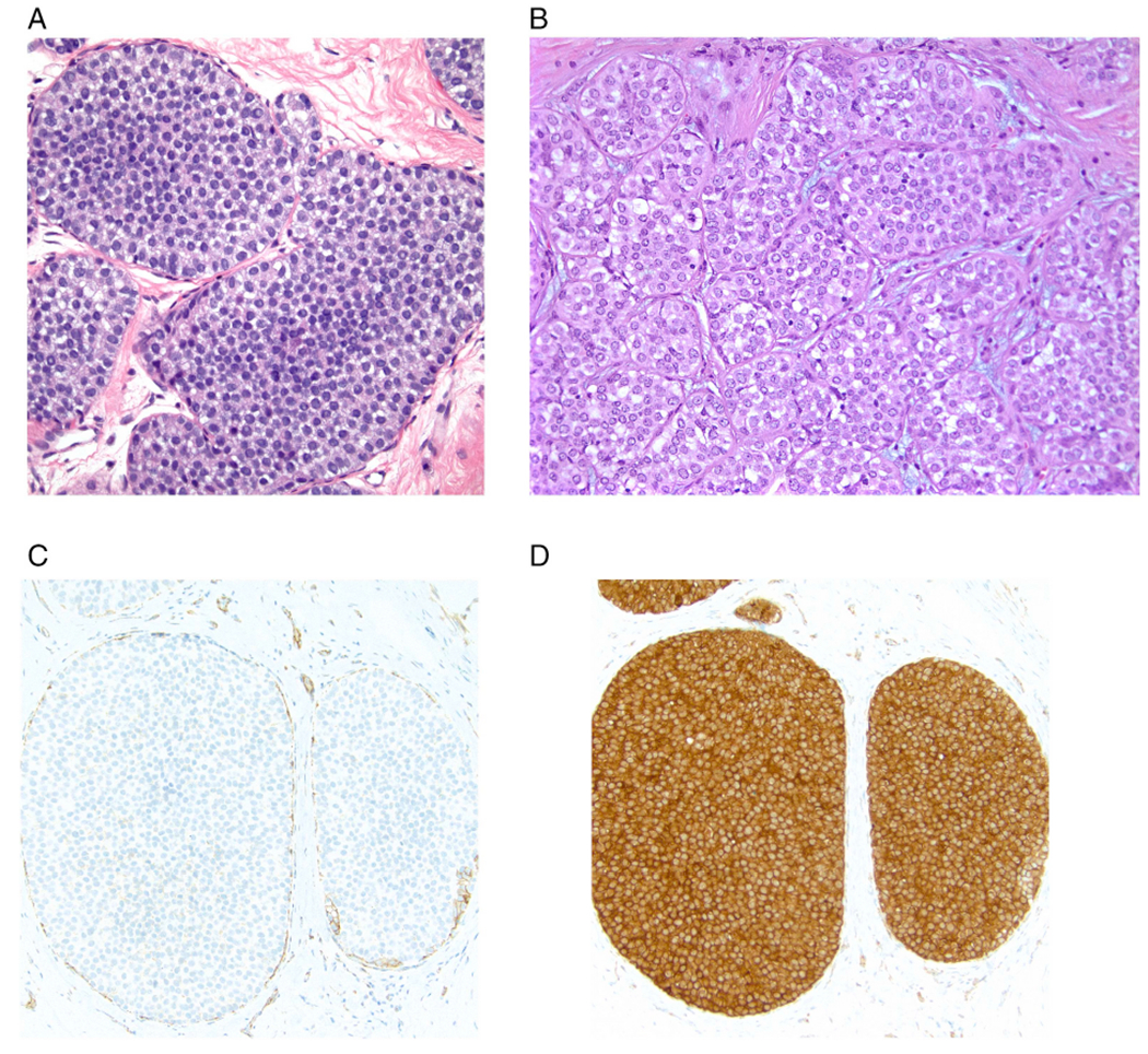
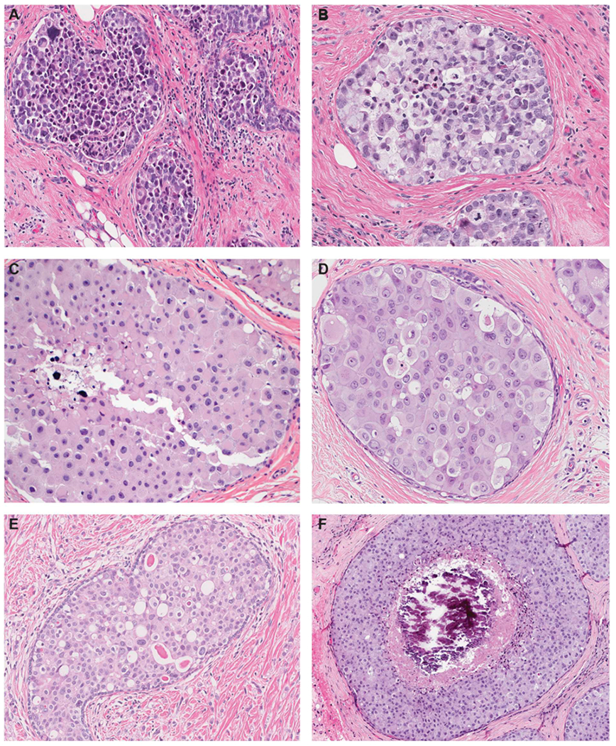
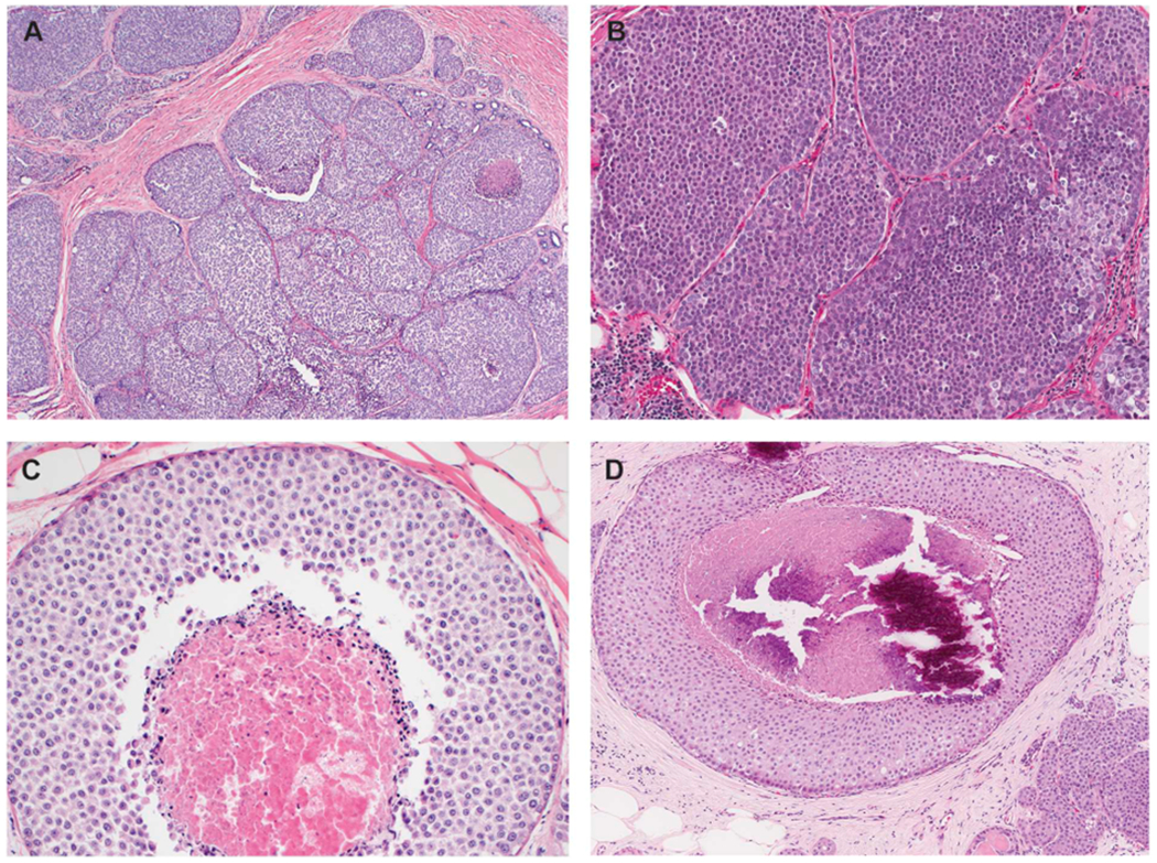
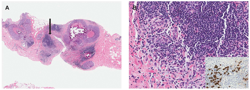
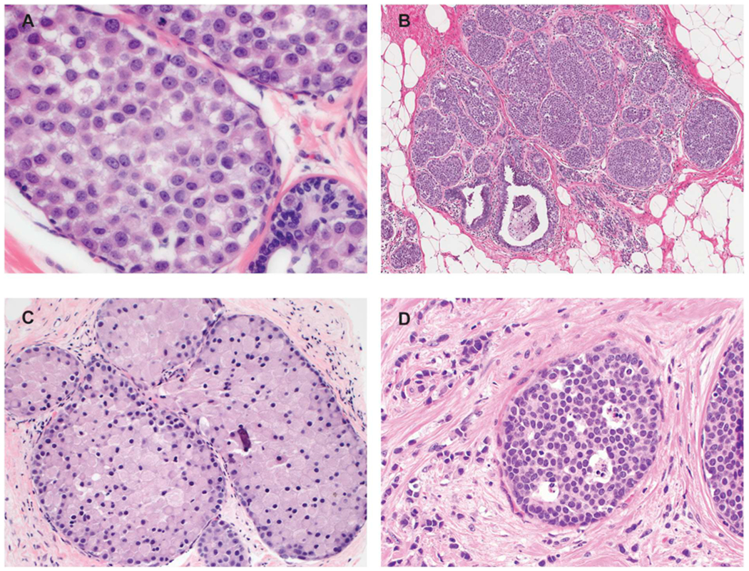
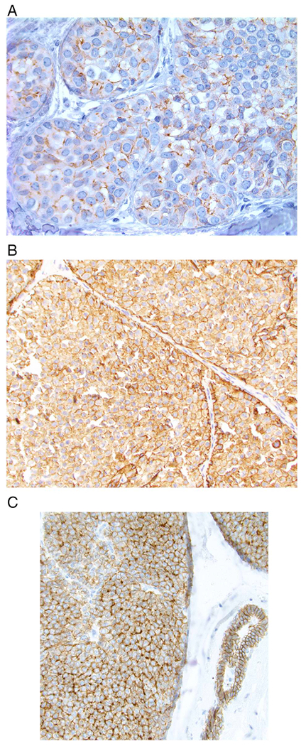
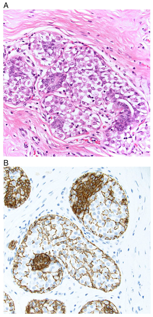
This review reflects a collaboration between the American Registry of Pathology (the publisher of the Armed Forces Institute of Pathology Fascicles) and Annals of Diagnostic Pathology. It is part of a series of expert recommendations on topics encountered in daily practice. The authors, 4 pathologists with expertise in breast pathology and a breast surgeon with a clinical and research interest in lobular carcinoma in situ (LCIS), met by conference call in September 2019 to develop recommendations for evaluating and reporting LCIS. Herein, we summarize the diagnostic criteria of classic LCIS and LCIS subtypes according to the most recent WHO criteria, discuss how best to distinguish LCIS from ductal carcinoma in situ in problematic cases (including the uses and limitations of E-cadherin immunohistochemistry), and review outcome and management issues for patients with LCIS.


Figures

Fig. 1.
Fig. 1.
Classic lobular carcinoma in situ (LCIS). A. Type A cells. Small cells with uniform nuclei. B. Type B cells. The nuclei are larger and slightly more variable in size and shape than those seen in Type A cells. The chromatin is vesicular and small nucleoli are evident in some of the nuclei. C. E-cadherin immunostain demonstrating loss of membrane expression in the LCIS cells (note staining of surrounding myoepithelial cells). D. p120 catenin immunostain showing cytoplasmic staining of the LCIS cells.
Fig. 2.
Fig. 2.
Pleomorphic lobular carcinoma in situ (LCIS). A. Pleomorphic LCIS is characterized by a solid proliferation of dyscohesive cells, similar to classic LCIS, but with marked nuclear pleomorphism equivalent to high-grade DCIS. B. In this pleomorphic LCIS, many neoplastic cells have signet-ring cell morphology with large intracytoplasmic mucin-filled vacuoles that displace the nuclei to the periphery. Also noted are multinucleated cells, which may be present in pleomorphic LCIS but are not observed in classic LCIS. C and D. A subset of pleomorphic LCIS have apocrine features and are characterized by large cells with abundant eosinophilic, granular cytoplasm and large rounded nuclei with prominent nucleoli. E. Many neoplastic cells in this example of pleomorphic LCIS demonstrate prominent intracytoplasmic vacuoles, some of which contain a large eosinophilic globule. The finding of prominent intracytoplasmic vacuoles in an in situ lesion favors LCIS over DCIS, but may also create the false impression of glandular structures mimicking DCIS. Note the presence of associated invasive lobular carcinoma in the background. F. Pleomorphic LCIS is often, but not always, associated with comedo necrosis and calcifications.
Fig. 3.
Fig. 3.
Florid lobular carcinoma in situ (LCIS). A. Florid LCIS has cytologic features identical to those of classic LCIS but is distinguished by marked distention of TDLUs or ducts, creating a confluent mass-like appearance at low power view. To qualify for florid subtype, an LCIS lesion should demonstrate at least one of the two architectural features depicted in B and C: (B) the spaces are expanded to a point that there is little to no intervening stroma between the markedly distended acini and ducts; (C) the expanded duct fills at least one high power field (an area equivalent to ~40–50 cells in diameter). Similar to pleomorphic LCIS, these lesions often demonstrate comedo necrosis. D. Florid LCIS with comedo necrosis and calcifications. Note the presence of classic LCIS with similar cytologic features at right lower corner, the presence of which should alert the pathologist the possibility of a LCIS subtype and not solid pattern DCIS.
Fig. 4.
Fig. 4.
Pleomorphic LCIS associated with subtle invasive lobular carcinoma. A. This core biopsy shows pleomorphic LCIS with comedo necrosis and calcifications and foci of chronic inflammation. B. High power evaluation of one of the inflammatory foci (highlighted by the arrow in panel A) reveals subtle invasive lobular carcinoma admixed with and masked by the lymphocytic infiltrate. A keratin immunostain (inset) is very useful in highlighting the invasive tumor cells.
Fig. 5.
Fig. 5.
Borderline and unusual LCIS lesions. A. In this LCIS case, the nuclei are larger than those of typical type B cells but fall short of the marked atypia required for pleomorphic LCIS. The lesion should be categorized as classic LCIS composed of type B cells. B. This LCIS lesion shows abundant proliferation with significantly expanded acini. However, the acini remain separate from one another with persistence of intervening interlobular stroma and none of the distended acini fill one high power field. The lesion does not meet the criteria for florid LCIS and should be classified as classic LCIS. C. This example of LCIS with apocrine features but lacking marked nuclear pleomorphism is challenging to classify. It should not be diagnosed as apocrine pleomorphic LCIS or simply “non-classic LCIS”. One suggested approach is to give the descriptive diagnosis “LCIS with apocrine features” with a comment in the report. D. This LCIS lesion is composed of classic lobular cells (type A) but shows single-cell apoptosis and minute foci of necrosis. In the absence of marked nuclear atypia or duct-lobular expansion, the lesion is best classified as classic LCIS.
Fig. 6.
Fig. 6.
Examples of aberrant E-cadherin expression in lobular carcinoma in situ. A. Most often, aberrant E-cadherin expression is characterized by weak, partial, fragmented, or beaded membrane staining. B. In some cases there is more extensive membranous staining as well as diffuse cytoplasmic staining. C. An example of aberrant E-cadherin expression in which some cells show a perinuclear, dot-like pattern of staining (note normal membranous expression of E-cadherin in the duct on the right side of the image).
Fig. 7.
Fig. 7.
A. In this example of lobular carcinoma in situ (LCIS), residual normal ductal epithelial cells are evident on the H and E-stained section. B. E-cadherin immunostain shows strong membranous staining of residual ductal epithelial cells as well as staining of myoepithelial cells and their processes that are present between the LCIS cells. The E-cadherin staining of these normal elements should not be mistaken for staining of the LCIS cells.

References

    1. Foote FW Jr, Stewart FW. Lobular carcinoma in situ: a rare form of mammary cancer. Am J Pathol 1941;17:419–99. - PMC - PubMed
    1. Dabbs DJ, Schnitt SJ, Geyer FC, Weigelt B, Baehner FL, Decker T, et al. Lobular neoplasia of the breast revisited with emphasis on the role of E-cadherin immunohistochemistry. Am J Surg Pathol 2013;37(7):e1–11. - PubMed
    1. Canas-Marques R, Schnitt SJ. E-cadherin immunohistochemistry in breast pathology: uses and pitfalls. Histopathology. 2016;68(1):57–69. - PubMed
    1. Chen YYDT, King TA, Palacios J, Shin SJ, Simpson PT. Lobular carcinoma in situ In: Board TWCE, editor. Breast Tumours. Lyon: International Agency for Research on Cancer; 2019. p. 71–4.
    1. Sneige N, Wang J, Baker BA, Krishnamurthy S, Middleton LP. Clinical, histopathologic, and biologic features of pleomorphic lobular (ductal-lobular) carcinoma in situ of the breast: a report of 24 cases. Mod Pathol 2002;15(10):1044–50. - PubMed

MeSH terms