. 2020 Feb 27;21(5):1619.
doi: 10.3390/ijms21051619.

A Facile One-Pot Synthesis of Versatile PEGylated Platinum Nanoflowers and Their Application in Radiation Therapy

Affiliations

A Facile One-Pot Synthesis of Versatile PEGylated Platinum Nanoflowers and Their Application in Radiation Therapy

Xiaomin Yang et al. Int J Mol Sci. .

Abstract

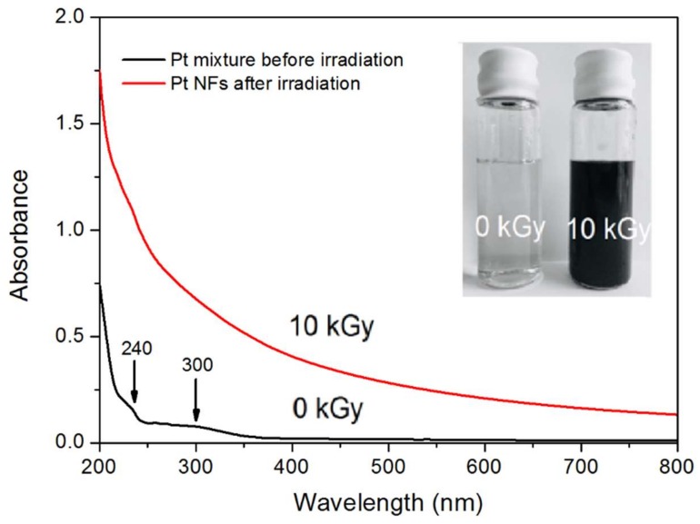
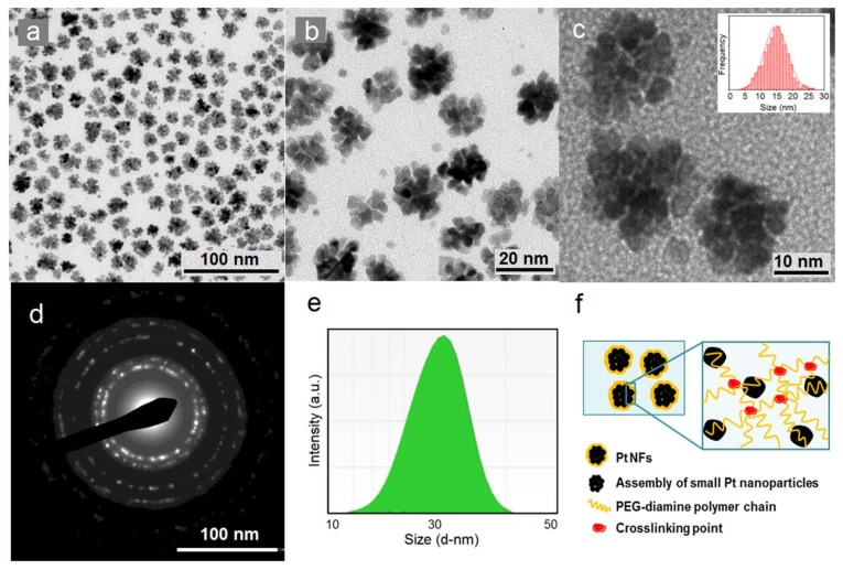
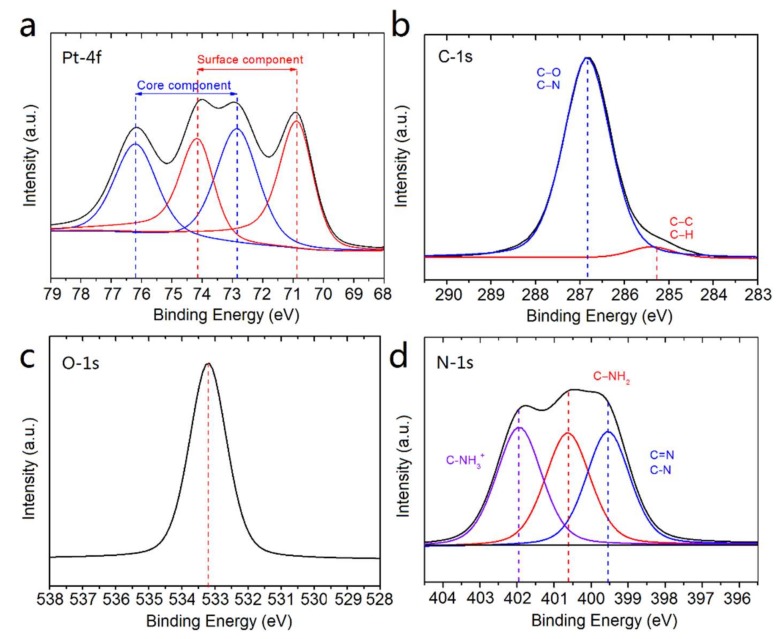
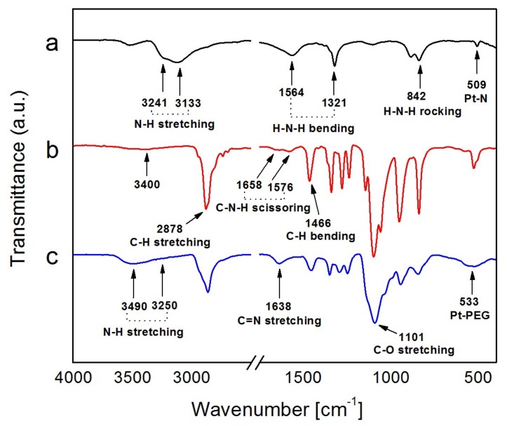
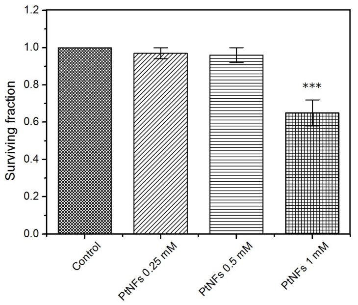
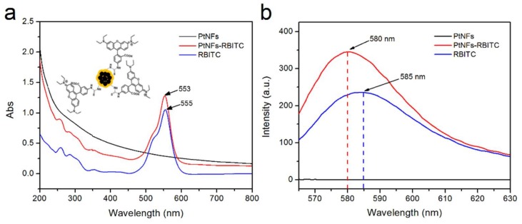
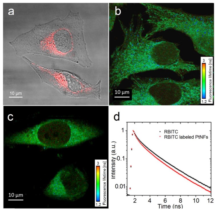
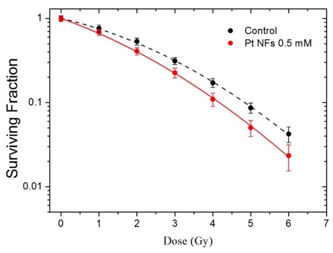
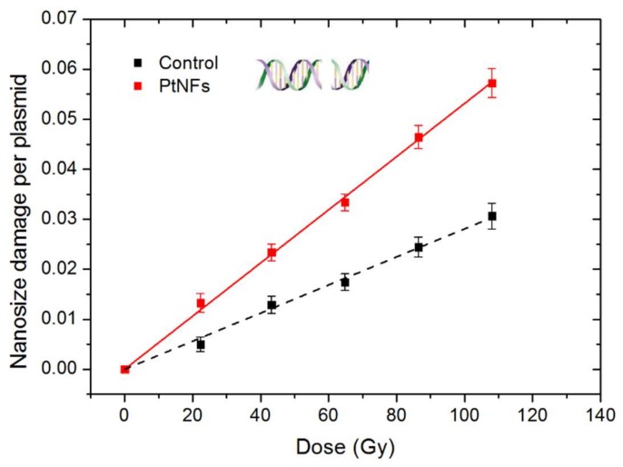
Nanomedicine has stepped into the spotlight of radiation therapy over the last two decades. Nanoparticles (NPs), especially metallic NPs, can potentiate radiotherapy by specific accumulation into tumors, thus enhancing the efficacy while alleviating the toxicity of radiotherapy. Water radiolysis is a simple, fast and environmentally-friendly method to prepare highly controllable metallic nanoparticles in large scale. In this study, we used this method to prepare biocompatible PEGylated (with Poly(Ethylene Glycol) diamine) platinum nanoflowers (Pt NFs). These nanoagents provide unique surface chemistry, which allows functionalization with various molecules such as fluorescent markers, drugs or radionuclides. The Pt NFs were produced with a controlled aggregation of small Pt subunits through a combination of grafted polymers and radiation-induced polymer cross-linking. Confocal microscopy and fluorescence lifetime imaging microscopy revealed that Pt NFs were localized in the cytoplasm of cervical cancer cells (HeLa) but not in the nucleus. Clonogenic assays revealed that Pt NFs amplify the gamma rays induced killing of HeLa cells with a sensitizing enhancement ratio (SER) of 23%, thus making them promising candidates for future cancer radiation therapy. Furthermore, the efficiency of Pt NFs to induce nanoscopic biomolecular damage by interacting with gamma rays, was evaluated using plasmids as molecular probe. These findings show that the Pt NFs are efficient nano-radio-enhancers. Finally, these NFs could be used to improve not only the performances of radiation therapy treatments but also drug delivery and/or diagnosis when functionalized with various molecules.

Keywords: cancer treatment; nanoparticle; platinum; radiation; radioenhancement; radiolysis; radiosensitization.


Conflict of interest statement

The authors declare no conflict of interest. The funders had no role in the design of the study; in the collection, analyses, or interpretation of data; in the writing of the manuscript, or in the decision to publish the results.

Figures

Figure 1
Figure 1
UV-Vis absorption spectra of diluted Pt containing solutions (10−3 mol L−1) before (black line) and after (red line) irradiation (optical path = 2 mm). Inserts: images of solutions resulting from 0 and 10 kGy radiation doses.
Figure 2
Figure 2
(ac) Lower and higher magnification High-Resolution Transmission Electron Microscopy (HR-TEM) images of platinum nanoflowers (Pt NFs) and HR-TEM size distribution histogram of Pt NFs (inset); (d) Electron diffraction pattern of Pt NFs; (e) Dynamic Light Scattering (DLS) size distribution by intensity of Pt NFs; (f) Scheme of the formation of Pt NFs by radiation-induced polymer cross-linking.
Figure 3
Figure 3
X-ray Photoelectron Spectroscopy (XPS) spectra of Pt NFs at (a) Pt-4f, (b) C-1s, (c) O-1s, (d) N-1s regions.
Figure 4
Figure 4
Fourier Transform InfraRed (FTIR) spectra of (a) Pt(NH3)4Cl2, (b) PEG-diamine 2000 and (c) Pt NFs.
Figure 5
Figure 5
Cytotoxicity determined by clonogenic assay following a 6 h exposure to medium containing Pt NFs at different Pt concentrations of 2.5 × 10⁻⁴, 5 × 10⁻⁴ and 10−3 mol L−1. Data represent the mean ± SD of three identical experiments made in triplicate. Asterisks denote significant differences with control cells from ANOVA: *** p ˂ 0.001.
Figure 6
Figure 6
(a) UV-Vis absorbance spectra of Pt NFs (black spectrum), Rhodamine B Isothiocyanate (RBITC) labeled Pt NFs (Red spectrum), free RhB-ITC (blue spectrum). Insert shows the schematic illustration of RBITC labeled Pt NFs. (b) Fluorescence emission spectra (λexc = 555 nm) of Pt NFs, RBITC labeled Pt NFs, RBITC.
Figure 7
Figure 7
(a) Merged image of the transmission and fluorescence images obtained by confocal microscopy of HeLa cell loaded with RBITC labeled Pt NFs at a concentration of Pt of 5 × 10⁻⁴ mol L−1, incubated for 6 h; (b,c) The Fluorescence Lifetime Imaging Microscopy (FLIM) imaging of cervical cancer cells (HeLa) incubated with RBITC labeled Pt NFs and free RBITC respectively, fluorescence lifetime is showed in the nanosecond range; (d) Fluorescence decay curves (mean lifetime curve) of the free RBITC (black) and of the RBITC labeled Pt NFs (red) after an excitation at 550 nm.
Figure 8
Figure 8
Surviving Fraction (SF) of HeLa cells irradiated by γ rays, in the presence of Pt NFs (red) and in the control (black).
Figure 9
Figure 9
Nanosize damage induced by γ-rays in plasmids loaded with Pt NFs (red solid line) and in the control (black dash line) as functions of the irradiation dose.

References

    1. Herold D.M., Stobbe C.C., Iyer R.V., Chapman J.D. Gold microspheres: A selective technique for producing biologically effective dose enhancement. Int. J. Radiat. Biol. 2000;76:1357–1364. - PubMed
    1. Kuncic Z., Lacombe S. Nanoparticle radio-enhancement: Principles, progress and application to cancer treatment. Phys. Med. Biol. 2018;63:02TR01. doi: 10.1088/1361-6560/aa99ce. - DOI - PubMed
    1. Hainfeld J.F., Dilmanian F.A., Slatkin D.N., Smilowitz H.M. Radiotherapy enhancement with gold nanoparticles. J. Pharm. Pharmacol. 2008;60:977–985. doi: 10.1211/jpp.60.8.0005. - DOI - PubMed
    1. Hainfeld J.F., Slatkin D.N., Smilowitz H.M. The use of gold nanoparticles to enhance radiotherapy in mice. Phys. Med. Biol. 2004;49:N309. doi: 10.1088/0031-9155/49/18/N03. - DOI - PubMed
    1. Haume K., Rosa S., Grellet S., Śmiałek M.A., Butterworth K.T., Solov’yov A.V., Prise K.M., Golding J., Mason N.J. Gold nanoparticles for cancer radiotherapy: A review. Cancer Nanotechnol. 2016;7:8. doi: 10.1186/s12645-016-0021-x. - DOI - PMC - PubMed