Review
. 2020 Feb 27;9(3):561.
doi: 10.3390/cells9030561.

Myeloid-Derived Suppressor Cells as a Therapeutic Target for Cancer

Affiliations
Review

Myeloid-Derived Suppressor Cells as a Therapeutic Target for Cancer

Andrew M K Law et al. Cells. .

Abstract

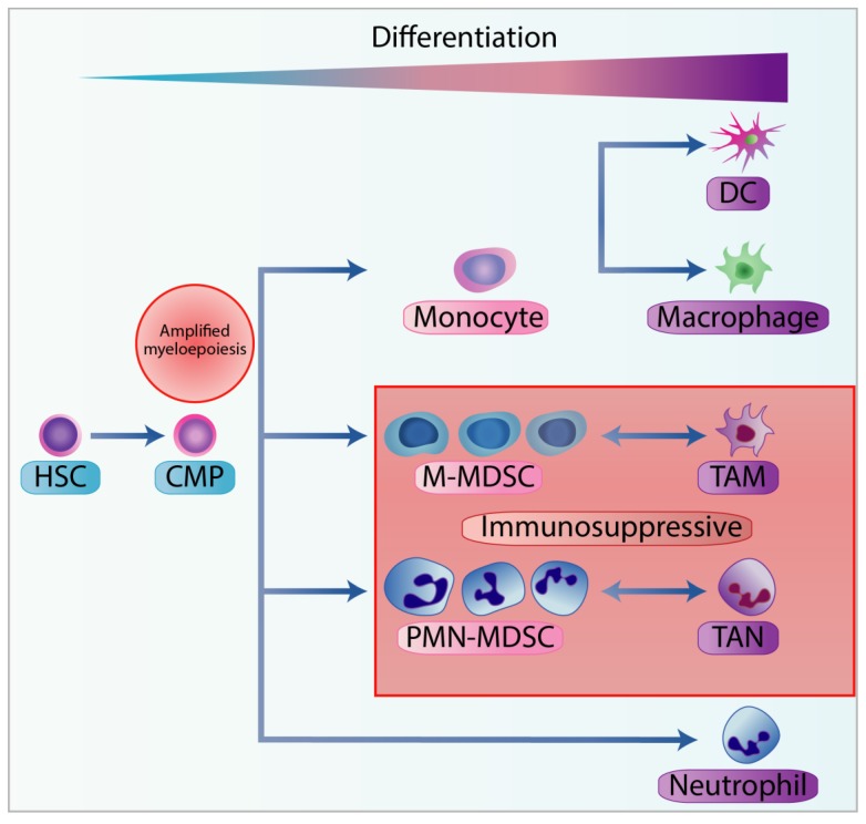
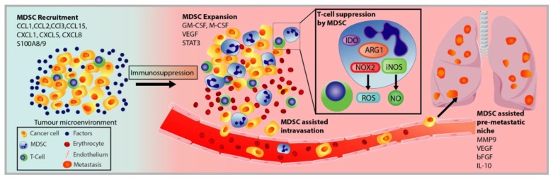
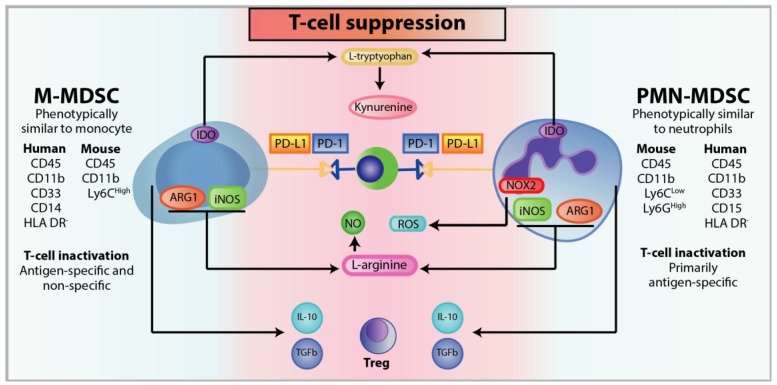
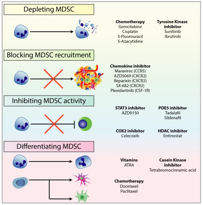
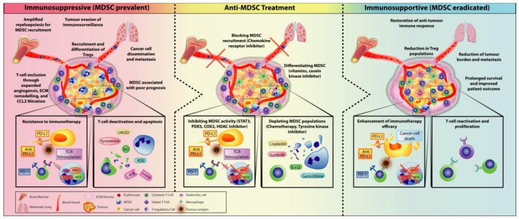
The emergence of immunotherapy has been an astounding breakthrough in cancer treatments. In particular, immune checkpoint inhibitors, targeting PD-1 and CTLA-4, have shown remarkable therapeutic outcomes. However, response rates from immunotherapy have been reported to be varied, with some having pronounced success and others with minimal to no clinical benefit. An important aspect associated with this discrepancy in patient response is the immune-suppressive effects elicited by the tumour microenvironment (TME). Immune suppression plays a pivotal role in regulating cancer progression, metastasis, and reducing immunotherapy success. Most notably, myeloid-derived suppressor cells (MDSC), a heterogeneous population of immature myeloid cells, have potent mechanisms to inhibit T-cell and NK-cell activity to promote tumour growth, development of the pre-metastatic niche, and contribute to resistance to immunotherapy. Accumulating research indicates that MDSC can be a therapeutic target to alleviate their pro-tumourigenic functions and immunosuppressive activities to bolster the efficacy of checkpoint inhibitors. In this review, we provide an overview of the general immunotherapeutic approaches and discuss the characterisation, expansion, and activities of MDSCs with the current treatments used to target them either as a single therapeutic target or synergistically in combination with immunotherapy.

Keywords: Myeloid derived suppressor cells; immune checkpoint inhibitors; immune system; immunotherapy; tumour microenvironment.


Conflict of interest statement

The authors declare no conflict of interest.

Figures

Figure 1
Figure 1
Workflow of adoptive T-cell therapy using TILs or receptor modified T-cells. Adoptive T-cell therapy can improve anti-tumour response by expanding TIL populations extracted from patient tumour (left), or genetically modifying the TCR or generating a chimeric antigen receptor (CAR) (right). With TIL expansion, the patient tumour is surgically resected and the TILs are isolated and expanded ex vivo. The TIL populations are then further increased through a Rapid Expansion Protocol before they are intravenously infused back into the lymphodepleted patient. For the genetic modification of T-cell, the TCR and CAR-T therapy extracts T-cells from the peripheral blood via leukapheresis and are transduced with viral vectors to express a modified TCR or CAR. In both approaches, the patient is lymphodepleted with cyclophosphamide before T-cell infusion and is administered with IL-2 to improve treatment efficacy and longevity.
Figure 2
Figure 2
Immune checkpoint blockade of T-cell activity and mechanism of action of checkpoint inhibitors. The immune checkpoints regulate T-cell activity and are crucial for maintaining self-tolerance. However, in cancer, the endogenous T-cell immune checkpoints, CTLA-4 and PD-1, inhibit T-cell activity when bound to their ligands, CD80/86 (antigen-presenting cells) and PD-L1 (cancer cells), respectively. Treatments with checkpoint inhibitors can disrupt this regulatory interaction allowing T-cell cytotoxic activity against cancer cells.
Figure 3
Figure 3
Stages of myelopoiesis differentiation in cancer. Myelopoiesis is amplified during chronic inflammation to assist tumour progression and dissemination. The hematopoietic stem cells (HSC) differentiate into the common myeloid progenitor (CMP), which can further differentiate through the hematopoietic system. In physiological conditions, CMP can differentiate into neutrophils or into monocytes, and subsequently into dendritic cells (DC) or macrophages. However, with chronic inflammation, pro-inflammatory cytokines can skew the monocytopoiesis of CMP into monocytic-myeloid-derived suppressor cells (M-MDSC) and tumour-associated macrophages (TAM), and granulopoiesis into polymorphonuclear myeloid-derived suppressor cells (PMN-MDSC) and tumour-associated neutrophils (TAN).
Figure 4
Figure 4
Schematic of MDSC recruitment and role in cancer progression and metastatic spread. MDSC are recruited to the tumour site by the same factors that mobilise neutrophils and monocytes. Within the tumour microenvironment, the MDSC population expands and exerts their immunosuppressive functions to induce T-cell and NK cell anergy through different mechanisms, such as through the enzymes IDO, ARG1, iNOS, and NOX2. MDSC can also assist in cancer cell dissemination through the promotion of angiogenesis, EMT and MET transition, and secretion of tumourigenic factors.
Figure 5
Figure 5
Mechanisms of T-cell suppression with phenotypic and functional differences between M-MDSC and PMN-MDSC. Both M-MDSC and PMN-MDSC display different cell surface markers and mechanisms for immunosuppression. Various mechanisms are used to suppress T-cell activity or induce T-cell apoptosis. (Top to bottom) L-tryptophan catabolism by IDO results in tryptophan starvation, leading to T-cell anergy, cell cycle arrest, and promotion of CD4 T-cells to differentiate into Tregs. Similarly, kynurenine, a tryptophan-derived catabolite by IDO inhibits T-cell and NK cell proliferation and promotes their apoptosis. In addition, kynurenine can bind to the aryl hydrocarbon receptor on T-cells to induce differentiation into Tregs. MDSCs can also induce T-cell exhaustion through elevated expression of PD-L1 to interact with the immune checkpoint PD-1. L-arginine is an essential amino acid that regulates T-cell cell cycle progression. Depletion of L-arginine by iNOS and ARG1 results in G0-G1 arrest in T-cells and downregulation of the TCR ζ-chain. The TCR will also undergo nitrosylation leading to impaired TCR signaling that is necessary for T-cell function. TCR nitrosylation results from high concentrations of NO, generated by iNOS catabolism of L-arginine, and ROS, a by-product of NOX2. MDSC can also recruit Tregs and induce their expansion via the secretion of cytokines such as IL-10 and TGFB.
Figure 6
Figure 6
Treatments used to target different mechanisms associated with pro-tumourigenic MDSC. There are multiple therapeutic approaches against MDSC to restore anti-tumour functions in immune cells and improve immunotherapy, in particular checkpoint inhibitors. These approaches include: (1) depleting MDSC populations through low-dose chemotherapy and tyrosine kinase inhibitors; (2) preventing MDSC recruitment to the TME by targeting chemokine receptors responsible for the recruitment and migration of MDSCs; (3) attenuating the immunosuppressive mechanisms of MDSC by downregulating the expression of ARG1 and iNOS, and reducing ROS generation; (4) inducing the differentiation of MDSC into mature myeloid cells to reduce MDSC population and remove their immunosuppression.
Figure 7
Figure 7
Treatment of MDSC to alleviate an immunosuppressive environment as an approach to enhancing immunotherapeutic treatments by shifting towards an immunosupportive TME. The immunosuppressive TME is propagated by various suppressive cells such as MDSCs and Tregs. Recruitment of MDSC within the TME can promote tumour expansion through various mechanisms (developing a pre-metastatic niche to help cancer cell metastasis, inducing resistance to immunotherapy by preventing the infiltration of T-cell into the tumour, suppressing and deactivating T-cell function, and inducing T-cell apoptosis) and recruitment of Tregs to further amplify immunosuppression. Thus, MDSC is often associated with poor prognosis in patients. Anti-MDSC treatments have become a major clinical target to re-establish immune control against cancer. By creating an immunosupportive environment, T-cell activity is restored, which leads to improved immunotherapy efficacy. Overall, this has resulted in prolonged survival and reduction of metastasis and tumour regression.

Similar articles

Cited by

References

    1. Garcia-Lora A., Algarra I., Garrido F. MHC class I antigens, immune surveillance, and tumor immune escape. J. Cell Physiol. 2003;195:346–355. doi: 10.1002/jcp.10290. - DOI - PubMed
    1. Fernald K., Kurokawa M. Evading apoptosis in cancer. Trends Cell Biol. 2013;23:620–633. doi: 10.1016/j.tcb.2013.07.006. - DOI - PMC - PubMed
    1. Burkholder B. Tumor-induced perturbations of cytokines and immune cell networks. Biochim. Biophys. Acta. 2014;1845:182–201. doi: 10.1016/j.bbcan.2014.01.004. - DOI - PubMed
    1. Salgado R. The evaluation of tumor-infiltrating lymphocytes (TILs) in breast cancer: Recommendations by an International TILs Working Group 2014. Ann. Oncol. 2015;26:259–271. doi: 10.1093/annonc/mdu450. - DOI - PMC - PubMed
    1. Chow M.T., Moller A., Smyth M.J. Inflammation and immune surveillance in cancer. Semin Cancer Biol. 2012;22:23–32. doi: 10.1016/j.semcancer.2011.12.004. - DOI - PubMed

Publication types

Substances