Review
. 2020 Feb 27;9(3):640.
doi: 10.3390/jcm9030640.

Histone Post-Translational Modifications and CircRNAs in Mouse and Human Spermatozoa: Potential Epigenetic Marks to Assess Human Sperm Quality

Affiliations
Review

Histone Post-Translational Modifications and CircRNAs in Mouse and Human Spermatozoa: Potential Epigenetic Marks to Assess Human Sperm Quality

Teresa Chioccarelli et al. J Clin Med. .

Abstract

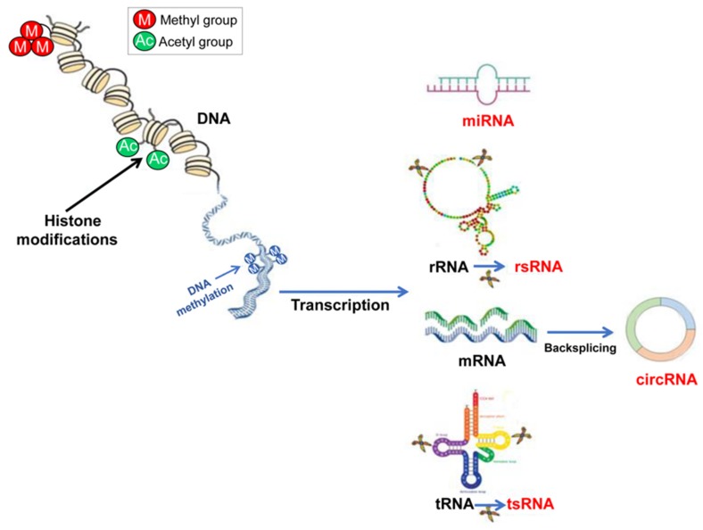
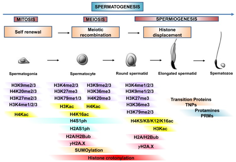
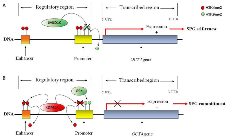
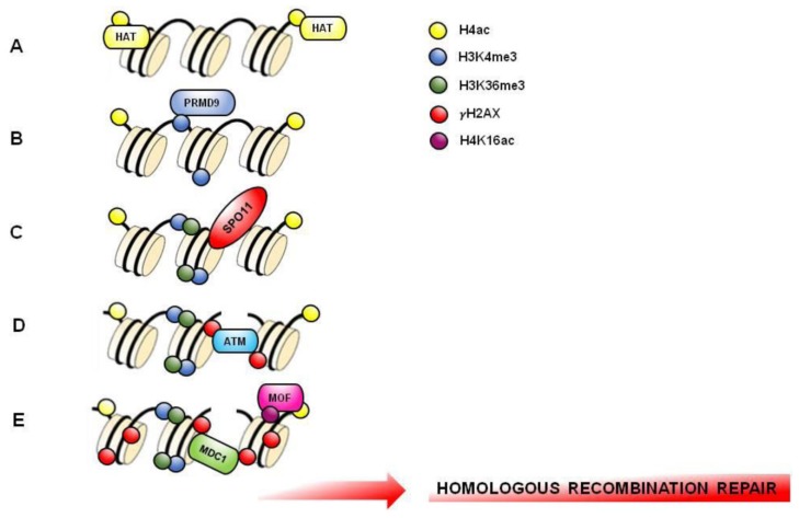
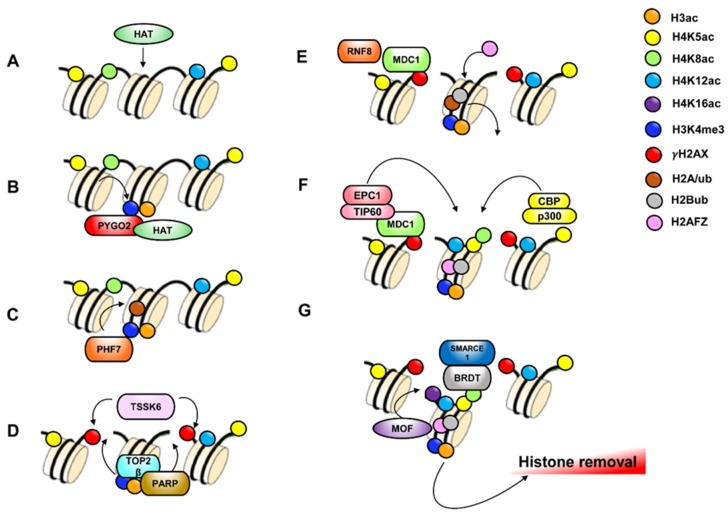
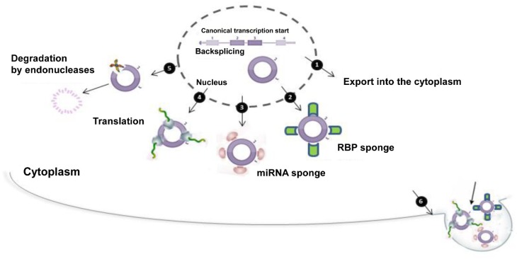
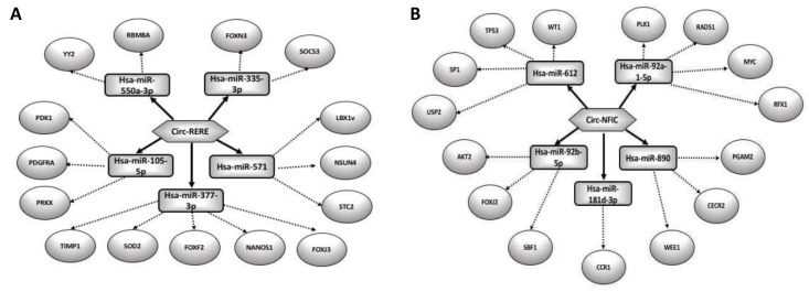
Spermatozoa (SPZ) are motile cells, characterized by a cargo of epigenetic information including histone post-translational modifications (histone PTMs) and non-coding RNAs. Specific histone PTMs are present in developing germ cells, with a key role in spermatogenic events such as self-renewal and commitment of spermatogonia (SPG), meiotic recombination, nuclear condensation in spermatids (SPT). Nuclear condensation is related to chromatin remodeling events and requires a massive histone-to-protamine exchange. After this event a small percentage of chromatin is condensed by histones and SPZ contain nucleoprotamines and a small fraction of nucleohistone chromatin carrying a landascape of histone PTMs. Circular RNAs (circRNAs), a new class of non-coding RNAs, characterized by a nonlinear back-spliced junction, able to play as microRNA (miRNA) sponges, protein scaffolds and translation templates, have been recently characterized in both human and mouse SPZ. Since their abundance in eukaryote tissues, it is challenging to deepen their biological function, especially in the field of reproduction. Here we review the critical role of histone PTMs in male germ cells and the profile of circRNAs in mouse and human SPZ. Furthermore, we discuss their suggested role as novel epigenetic biomarkers to assess sperm quality and improve artificial insemination procedure.

Keywords: circular RNAs (circRNAs); embryo development; fertilization; histone post-translational modifications (histone PTMs); human male infertility; sperm; spermatogenesis.


Conflict of interest statement

The authors declare no conflict of interest.

Figures

Figure 1
Figure 1
A schematic view of the main epigenetic processes. Gene expression can be epigenetically regulated at the transcriptional level, via DNA methylation and/or through histone histone PTMs able to conduct chromatin remodeling. The epigenetic regulation at the translational level is mainly under the control of ncRNAs. In sperm cells, recent results point to some classes of ncRNAs as mainly involved in their physiology, miRNAs, rsRNAs, tsRNAs and circRNAs, the last cited produced by mRNAs through a backsplicing reaction.
Figure 2
Figure 2
Histone PTMs regulate germ cell progression. The main histone PTMs involved in chromatin dynamic organization during proliferation, meiosis, and differentiation of germ cells, from spermatogonia to spermatozoa (spermatogenesis). In detail, histone PTMs during germ cell progression, self-renewal, meiotic recombination and histone-protamine transition.
Figure 3
Figure 3
Hypothetical molecular mechanism involved in OCT4 gene regulation in spermatogonia (SPG) stem cells. OCT4 gene is represented. (A) Active transcriptional mark H3K4me2 (red circle) at the promoter and proximal enhancer of OCT4 gene favors transcriptional activation; demethylation of repressive transcriptional mark H3K9me2 (green circle) by JMJD1C demethylase may participate to OCT4 gene activation, favoring SPG self-renewal. (B) H3K9me2, by G9a methylase, blocks OCT4 gene expression; H3K4me2 demethylation, by KDM1A demethylase, may participate to transcriptional repression of stem cell factor OCT4 favoring SPG commitment and differentiation.
Figure 4
Figure 4
Hypothetical molecular mechanism involved in meiotic hotspots formation. DNA and several nucleosomes are represented. (A) Histone H4 acetylation (H4K5ac, H4K8ac, H4K12ac, H4K16ac) (yellow circle) is enriched in PL-SPC to favor an opened chromatin structure that facilitate recombination hotspots formation. (B) PRDM9 binds DNA and catalyzes H3K4me3 formation (blue circle) on hotspots. (C) H3K36me3 (green circle) associates with H3K4me3 on the same nucleosome to create a hotspot-specific signature recognized by endonuclease SPO11. (D) DNA double strand breaks (DSBs) formed by SPO11 triggers the phosphorylation of histone H2AX (γH2AX; red circle) by ATM kinase. (E) MOF acetylase induces H4K16ac (purple circle) facilitating MDC1 recruitment. MDC1 binds γH2AX and amplifies its phosphorylation to induce the recruitment of DNA repair factors starting homologous recombination.
Figure 5
Figure 5
Hypothetical molecular mechanism involved in histone displacement. DNA and several nucleosomes are represented. (A) Histone H4 acetylation (green-yellow-light blue circles) catalyzed by Histone Acetyl Transferase (HAT) occurs to favor an opened chromatin structure that facilitate histone removal. (B) PYGO2 recognizes H3K4me3 (blue circle) and facilitate HAT recruitment to induce H3 acetylation (orange circle). (C) PHF7 recognizes H3K4me3 to induce H2Aub (brown circle). (D) TOP2β nuclease introduces DSBs and induces poly ADP-ribose polymerases (PARPs) activation. PARPs activity is required for TSSK6, a serine-protein kinase, that forms γH2AX. (E) MDC1 binds γH2AX and recruits ubiquitin E3 ligase RNF8 that catalyzes H2Bub (grey circle). H2Aub is replaced by histone variant H2AFZ (pink circle). (F) MDC1 recruits NuA4 HAT complex (EPC1-TIP60) that, in association with CBP-p300 HAT complex, induce H4 acetylation on K5, 8 and 12 (yellow-green-light blue circles). (G) Dimer H2AFZ/H2Bub facilitate association of MOF HAT to the chromatin for H4K16ac formation as last step of histone H4 hyperacetylation Finally, BRDT, in association to SMARCE1, binds K5ac and K8ac of tetra-acetylated H4 to guide histone removal.
Figure 6
Figure 6
CircRNAs at a glance. Starting from a canonical mRNA, circRNA biogenesis starts through a backsplicing reaction. This event takes place in the nucleus. After that, circRNA may be exported from the nucleus into the cytoplasm (1). In the cytoplasm, circRNA may have multiple actions: it is able to bind to RBPs (2) and/or miRNAs (3) with a typical “sponge activity”; if equipped with IRES sequences, it may be translated (4); circRNA degradation may be possible through endonuclease activity (5); the production of vescicles containing circRNA is a way to remove it from the cytoplasm. After its release into the extracellular space, it probably reaches other cells or tissues perhaps acting as a signal or playing other unknown functions (6).
Figure 7
Figure 7
CircRNA-miRNA network analysis. Circ-RERE and circ-NFIC and their predicted miRNAs were selected to generate a network map involved in embryo development (A) and germ cell progression (B), respectively. The circRNA-miRNA network was constructed using bioinformatic online programs (starBase, circBase, TargetScan, miRBase, Cytoscape).

References

    1. Holliday R. The inheritance of epigenetic defects. Science. 1987;238:163–170. doi: 10.1126/science.3310230. - DOI - PubMed
    1. Jenkins T.G., Carrell D.T. The sperm epigenome and potential implications for the developing embryo. Reproduction. 2012;143:727–734. doi: 10.1530/REP-11-0450. - DOI - PubMed
    1. Chianese R., Pierantoni R. Sperm Cells, an efficient shuttle for the intergenerational epigenetic memory. J. Pituit. Res. Treat. 2016;1:e001.
    1. Conine C., Sun F., Song L., Rivera-Pérez J.A., Rando O. Small RNAs Gained during Epididymal Transit of Sperm Are Essential for Embryonic Development in Mice. Dev. Cell. 2018;46:470–480. doi: 10.1016/j.devcel.2018.06.024. - DOI - PMC - PubMed
    1. Beaujean N. Histone post-translational modifications in preimplantation mouse embryos and their role in nuclear architecture. Mol. Reprod. Dev. 2013;81:100–112. doi: 10.1002/mrd.22268. - DOI - PubMed

LinkOut - more resources