. 2020 Feb 29;12(3):567.
doi: 10.3390/cancers12030567.

A Preclinical Embryonic Zebrafish Xenograft Model to Investigate CAR T Cells In Vivo

Affiliations

A Preclinical Embryonic Zebrafish Xenograft Model to Investigate CAR T Cells In Vivo

Susana Pascoal et al. Cancers (Basel). .

Abstract

Chimeric antigen receptor (CAR) T cells have proven to be a powerful cellular therapy for B cell malignancies. Massive efforts are now being undertaken to reproduce the high efficacy of CAR T cells in the treatment of other malignancies. Here, predictive preclinical model systems are important, and the current gold standard for preclinical evaluation of CAR T cells are mouse xenografts. However, mouse xenograft assays are expensive and slow. Therefore, an additional vertebrate in vivo assay would be beneficial to bridge the gap from in vitro to mouse xenografts. Here, we present a novel assay based on embryonic zebrafish xenografts to investigate CAR T cell-mediated killing of human cancer cells. Using a CD19-specific CAR and Nalm-6 leukemia cells, we show that live observation of killing of Nalm-6 cells by CAR T cells is possible in zebrafish embryos. Furthermore, we applied Fiji macros enabling automated quantification of Nalm-6 cells and CAR T cells over time. In conclusion, we provide a proof-of-principle study that embryonic zebrafish xenografts can be used to investigate CAR T cell-mediated killing of tumor cells. This assay is cost-effective, fast, and offers live imaging possibilities to directly investigate CAR T cell migration, engagement, and killing of effector cells.

Keywords: CAR T cells; CD19 CAR; in vivo imaging; zebrafish xenografts.


Conflict of interest statement

The authors declare no conflict of interest.

Figures

Figure 1
Figure 1
Chimeric antigen receptor (CAR) T cell mediated killing of Nalm-6 cells in vitro. (A) Efficacy of DiI-labeling in primary human T cells was analyzed by flow cytometry (excitation at 561 nm, detection with a 582/15 bandpass filter). Unlabeled T cells (“unstained”; filled grey histogram) served as a control. One representative experiment is shown. (B) Schematic of the CD19-specific second-generation CAR (FMC63.4-1BB.ζ) in which the FMC63-based scFv was fused to the hinge and transmembrane region of CD8α and the cytoplasmic domains of 4-1BB and CD3ζ. (C) Expression of the CD19-specific CAR in primary human T cells. Flow cytometric analysis using Protein L as a detection reagent. Mock T cells (“no CAR”; filled grey histogram) served as a negative control. One representative experiment of three independent experiments is shown. (D) Influence of DiI-labeling on the cytolytic activity of CAR T cells. Primary human T cells expressing either the CD19-specific CAR or no CAR and labeled or not labeled with DiI were co-cultured with CD19pos Nalm-6 cells. Experiments were performed with E/T (effector/target) ratios of 0.5, 1, 2, 4, and 8, as indicated. Cytolytic activity was quantified using a luciferase-based cytotoxicity assay. Data are expressed as means ± SD of one experiment with three donors. (E) Influence of the temperature on the cytolytic activity of CAR T cells. Primary human T cells expressing either the CD19-specific CAR or no CAR were co-cultured at 35 °C or 37 °C with GFPpos/CD19pos Nalm-6 cells at an E/T ratio of 1:1. Cytolytic activity was quantified by using a counting bead-based cytotoxicity assay. Data are expressed as individual data points (and their means) obtained from one experiment with three different T cell donors.
Figure 2
Figure 2
Nalm-6 xenografts in zebrafish embryos. GFP-expressing Nalm-6 cells (green) were injected into zebrafish embryos around 48 hours post fertilization (hpf). (A) An image was recorded at approximately 2 hours post injection (hpi) and again at 24 hpi (A’). (B) Immunostaining of the tail region using an anti Ki67 antibody (red) at 24 hpi, (B’) magnification of a region in B showing Ki67 positive Nalm-6 cells (arrows). (C) Immunostaining for apoptotic cells using an antibody against active Caspase 3 (red), (C’) magnification of a region in C. The arrow indicates a cell with active Caspase 3. Images in (A) were recorded on a Zeiss Axio Zoom.V16 fluorescence stereo zoom microscope, and in (B) and (C) on a confocal Leica SP8 WLL microscope. Images were rendered with Adobe Photoshop CS6. The scale bar in (A) represents 500 µm, in (B and C) 75 µm, and in (B’ and C’) 25 µm.
Figure 3
Figure 3
CAR T cell-mediated killing of Nalm-6 cells in zebrafish. Zebrafish embryos were injected with Nalm-6 cells (green) at approximately 48 hpf. Around 2 hours later, either mock T cells (without a CAR) (red cells in (A)) or CD19 CAR T cells (red cells in (B)) were injected and images were recorded within 2 hours and again at 24 hours post injection of T cells. (C) The numbers of Nalm-6 cells and T cells were quantified at 2 hpi and 24 hpi based on the fluorescent area covered by cells in the tail (red box in B) using Fiji. The two time points are connected by a line for each embryo. From left to right: Nalm-6 cells in zebrafish without any T cells; Nalm-6 cells (in zebrafish with CD19 CAR T cells), CD19 CAR T cells (in zebrafish with Nalm-6); Nalm-6 cells (in zebrafish with mock T cells), mock T cells (in zebrafish with Nalm-6) at 2 hpi and 24 hpi. (D) Violin plots normalized to the area covered by fluorescent cells at 2 hpi revealing the change in distribution at 24 hpi. Nalm-6 cells together with mock T cells or alone without T cells show similar distributions at 24 hpi, whereas Nalm-6 injected with CD19 CAR T cells show a reduction compared to Nalm-6 alone or Nalm-6 with mock T cells. Images were recorded on a Zeiss Axio Zoom.V16 fluorescence stereo zoom microscope and rendered with Adobe Photoshop CS6. Scale bar in (A) represents 1 mm.
Figure 4
Figure 4
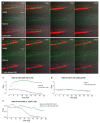
Quantifying specific elimination of Nalm-6 cells over time. Zebrafish embryos were injected with Nalm-6 cells (green) at approximately 48 hpf. Around 2 hours later, either CD19 CAR T cells (red cells in (A)) or T cells without a CAR (red cells in (B)) were injected and a time-lapse movie was recorded, starting approximately 4 hours post injection of T cells. Several time points of one embryo co-injected with CD19 CAR T cells (A) or mock T cells (B) are shown as single frames. (C) Quantification of cells over time based on the fluorescent area using Fiji. Nalm-6 (blue dots) in the presence of CD19 CAR T cells (shown as green dots), or the presence of T cells without CAR (D). (E) Nalm-6 in the presence of CD19 CAR T cells (blue) and T cells without CAR (green) over time. Images were recorded on a Leica SP8 X WLL confocal microscope and rendered with Adobe Photoshop CS6. Scale bar in (A) represents 250 µm.

References

    1. June C.H., O’Connor R.S., Kawalekar O.U., Ghassemi S., Milone M.C. CAR T cell immunotherapy for human cancer. Science. 2018;359:1361–1365. doi: 10.1126/science.aar6711. - DOI - PubMed
    1. June C.H., Sadelain M. Chimeric Antigen Receptor Therapy. N. Engl. J. Med. 2018;379:64–73. doi: 10.1056/NEJMra1706169. - DOI - PMC - PubMed
    1. Maude S.L., Laetsch T.W., Buechner J., Rives S., Boyer M., Bittencourt H., Bader P., Verneris M.R., Stefanski H.E., Myers G.D., et al. Tisagenlecleucel in Children and Young Adults with B-Cell Lymphoblastic Leukemia. N. Engl. J. Med. 2018;378:439–448. doi: 10.1056/NEJMoa1709866. - DOI - PMC - PubMed
    1. Labanieh L., Majzner R.G., Mackall C.L. Programming CAR-T cells to kill cancer. Nat. Biomed. Eng. 2018;2:377–391. doi: 10.1038/s41551-018-0235-9. - DOI - PubMed
    1. Mestermann K., Giavridis T., Weber J., Rydzek J., Frenz S., Nerreter T., Mades A., Sadelain M., Einsele H., Hudecek M. The tyrosine kinase inhibitor dasatinib acts as a pharmacologic on/off switch for CAR T cells. Sci. Transl. Med. 2019;11 doi: 10.1126/scitranslmed.aau5907. - DOI - PMC - PubMed

LinkOut - more resources