

Theoretical and Experimental Gas Volume Quantification of Micro- and Nanobubble Ultrasound Contrast Agents
- PMID: 32121484
- PMCID: PMC7150797
- DOI: 10.3390/pharmaceutics12030208
Theoretical and Experimental Gas Volume Quantification of Micro- and Nanobubble Ultrasound Contrast Agents
Abstract
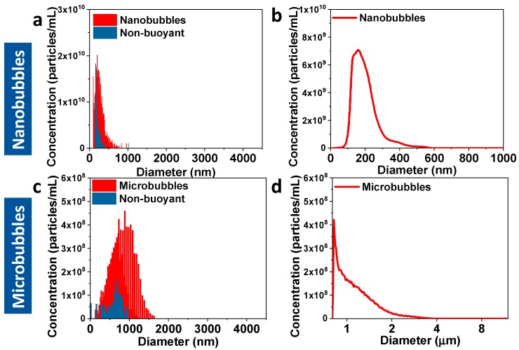
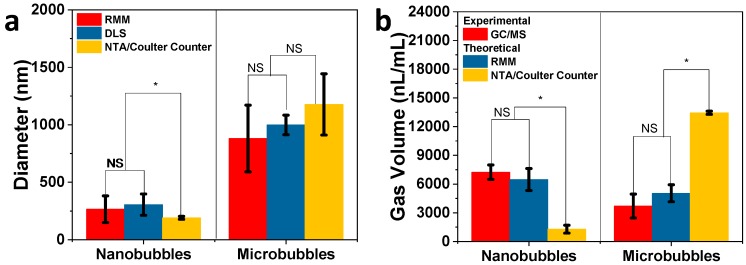
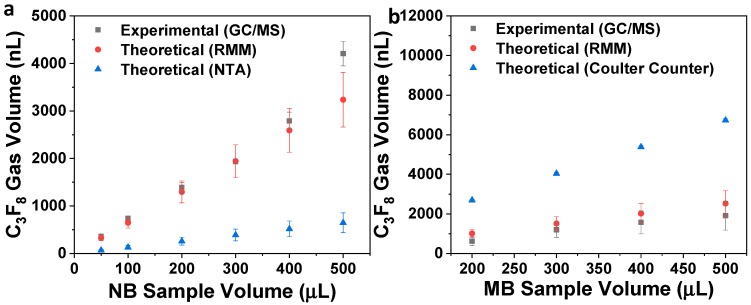
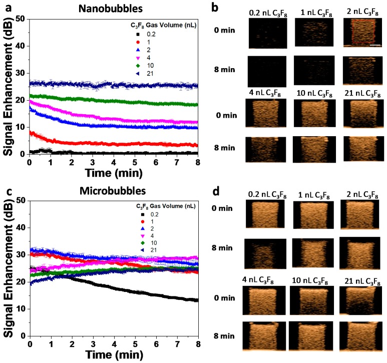
The amount of gas in ultrasound contrast agents is related to their acoustic activity. Because of this relationship, gas volume has been used as a key variable in normalizing the in vitro and in vivo acoustic behavior of lipid shell-stabilized bubbles with different sizes and shell components. Despite its importance, bubble gas volume has typically only been theoretically calculated based on bubble size and concentration that is typically measured using the Coulter counter for microbubbles and nanoparticle tracking analysis (NTA) for nanoscale bubbles. However, while these methods have been validated for the analysis of liquid or solid particles, their application in bubble analysis has not been rigorously studied. We have previously shown that resonant mass measurement (RMM) may be a better-suited technique for sub-micron bubble analysis, as it can measure both buoyant and non-buoyant particle size and concentration. Here, we provide validation of RMM bubble analysis by using headspace gas chromatography/mass spectrometry (GC/MS) to experimentally measure the gas volume of the bubble samples. This measurement was then used as ground truth to test the accuracy of theoretical gas volume predictions based on RMM, NTA (for nanobubbles), and Coulter counter (for microbubbles) measurements. The results show that the headspace GC/MS gas volume measurements agreed well with the theoretical predictions for the RMM of nanobubbles but not NTA. For nanobubbles , the theoretical gas volume using RMM was 10% lower than the experimental GC/MS measurements; meanwhile, using NTA resulted in an 82% lower predicted gas volume. For microbubbles, the experimental gas volume from the GC/MS measurements was 27% lower compared to RMM and 72% less compared to the Coulter counter results. This study demonstrates that the gas volume of nanobubbles and microbubbles can be reliably measured using headspace GC/MS to validate bubble size measurement techniques. We also conclude that the accuracy of theoretical predictions is highly dependent on proper size and concentration measurements.
Keywords: contrast agents; coulter counter; dynamic light scattering; gas chromatography/mass spectrometry; gas volume; microbubble; nanobubble; nanoparticle tracking analysis; perfluoropropane; resonant mass measurement; ultrasound.
Conflict of interest statement
The authors declare no conflict of interest. The funders had no role in the design of the study; in the collection, analyses, or interpretation of data; in the writing of the manuscript, or in the decision to publish the results.
Figures
References
-
- de Leon A., Perera R., Nittayacharn P., Cooley M., Jung O., Exner A.A. Ultrasound Contrast Agents and Delivery Systems in Cancer Detection and Therapy. Adv. Cancer Res. 2018;139:57–84. - PubMed
-
- American Society of Echocardiography (ASE) The Basics. [(accessed on 25 August 2019)]; Available online: https://www.asecho.org/contrast-zone/the-basics/
Grants and funding
LinkOut - more resources
Full Text Sources
Miscellaneous
