. 2020 Mar 2;147(5):dev184564.
doi: 10.1242/dev.184564.

Control of tissue morphogenesis by the HOX gene Ultrabithorax

Affiliations

Control of tissue morphogenesis by the HOX gene Ultrabithorax

Maria-Del-Carmen Diaz-de-la-Loza et al. Development. .

Abstract

Mutations in the Ultrabithorax (Ubx) gene cause homeotic transformation of the normally two-winged Drosophila into a four-winged mutant fly. Ubx encodes a HOX family transcription factor that specifies segment identity, including transformation of the second set of wings into rudimentary halteres. Ubx is known to control the expression of many genes that regulate tissue growth and patterning, but how it regulates tissue morphogenesis to reshape the wing into a haltere is still unclear. Here, we show that Ubx acts by repressing the expression of two genes in the haltere, Stubble and Notopleural, both of which encode transmembrane proteases that remodel the apical extracellular matrix to promote wing morphogenesis. In addition, Ubx induces expression of the Tissue inhibitor of metalloproteases in the haltere, which prevents the basal extracellular matrix remodelling necessary for wing morphogenesis. Our results provide a long-awaited explanation for how Ubx controls morphogenetic transformation.

Keywords: Development; Drosophila; Morphogenesis; Notopleural; Stubble.


Conflict of interest statement

Competing interestsThe authors declare no competing or financial interests.

Figures

Fig. 1.
Fig. 1.
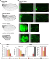
Ubx is required to repress expression of the Sb and Np aECM proteases in the haltere. (A) Maximum projection (Z-proj) of endogenous Stubble-GFP (Sb-GFP; top) and Notopleural-GFP (Np-GFP; bottom) localisation in developing wings at third instar larvae (L3), 4 h and 7 h APF. Wing hinge (h) and pouch (p) regions in L3 wing discs are indicated. Sb and Np start to be expressed at the end of the larval stage, mostly visible at the hinge folds. At 4 h APF, before ECM degradation, they strongly localise at the apical membrane of the wing epithelium, and their levels decrease in 7 h APF wings, which have already expanded and elongated after the ECM is degraded. Actin cytoskeleton is shown in red. (B) Quantification of Sb-GFP and Np-GFP immunofluorescence signal in the wing and the haltere at 4 h and 7 h APF. Both proteins are strongly expressed in the wing compared with the haltere. Wings show a maximum of GFP fluorescence at 4 h APF, whereas haltere fluorescence levels remain low. (C) Maximum projection (Z-proj) of Sb-GFP and Np-GFP localisation in developing halteres from third instar larvae (L3), at 4 h and 7 h APF. Halteres show consistently low levels of Np-GFP and Sb-GFP during all developmental stages. Actin cytoskeleton is shown in red. (D) Maximum projections of Sb-GFP (top) and Np-GFP (bottom) in ubxRNAi-expressing halteres (ubx-Gal4>UAS.UbxRNAi) at 4 h and 7 h APF. Loss of Ubx restores high levels of Sb and Np in the haltere at 4 h APF, leading to ectopic ECM degradation, which results in flattened and expanded halteres at 7 h APF. Dashed lines indicate the perimeter of the haltere, determined by looking at the actin cytoskeleton. (E) Quantification of Sb-GFP and Np-GFP immunofluorescence signal in control and ubx-Gal4>UAS.UbxRNAi wings and halteres at 4 h APF. Depletion of Ubx increases Sb-GFP and Np-GFP expression in the haltere to similar levels to the wing. Data are mean±s.d., n>4 for each developmental stage. *P<0.05, **P<0.005, ***P<0.001 (two-tailed Student's t-test). Scale bars: 50 μm.
Fig. 2.
Fig. 2.
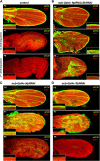
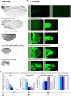
Depletion of Sb and Np impairs aECM remodelling and wing morphogenesis. (A) Adult wings from control animals and from animals with depletion of Np (nub-Gal4>NpRNAi), Sb (nub-Gal4>SbRNAi) or both (nub-Gal4>NpRNAi;SbRNAi). Simultaneous depletion of Sb and Np apical proteases during metamorphosis results in smaller and rounded wings. (B) Maximum projections of 7 h APF wings in control and mutant conditions with low levels of Np and Sb expressing Dp-YFP (aECM) or Vkg-GFP (bECM). Np depletion does not affect aECM degradation, and wings have elongated and expanded normally at 7 h APF; however, depleting Sb strongly impairs aECM degradation and wing expansion at 7 h APF, consistent with the strong expression of Sb at this stage of development. As loss of both Sb and Np is required to affect the adult wing, Np must function after 7 h APF to degrade the aECM, even in the absence of Sb (see Figs S1 and S2). Note that bECM degradation is not affected by depletion of apical proteases. Dashed lines indicate the perimeter of the wing blade, determined by looking at the actin cytoskeleton. (C) Quantification of size (area) and shape characteristics (aspect ratio, dorsoventral adhesion and epithelial folding) in control (w), nub-Gal4>NpRNAi, nub-Gal4>SbRNAi and nub-Gal4>NpRNAi;SbRNAi wings compared with control halteres (h). Mean±s.d. are shown from up to 20 wings or halteres for each genotype. Inhibition of aECM degradation by depletion of apical proteases decreases wing area and elongation, and impairs the adhesion of the dorsoventral layers, all features present in the haltere. aECM depletion also results in folding of the wing blade. Scale bars: 50 μm.
Fig. 3.
Fig. 3.
Apical ECM is degraded at late metamorphosis by Sb and Np. (A) Dumpy-YFP (Dp-YFP)-labelled aECM is normally degraded during pupal days P7 and P8. Actin is shown in red. (B) Silencing of both Sb and Np expression by RNAi with nub-Gal4 prevents aECM degradation at P7 and P8. (C) Silencing of Np expression alone by RNAi with nub-Gal4 prevents aECM degradation at P7 but not P8. (D) Silencing of Sb expression alone by RNAi with nub-Gal4 prevents aECM degradation at P7 but not P8. Insets show high magnification views of the pupal wing margin. Note the proximity of the wing margin bristles to the aECM in the control. Scale bars: 50 µm.
Fig. 4.
Fig. 4.
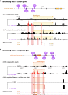
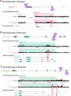
Ubx binds to specific sites in the Sb and Np genes in the haltere. (A) Ubx binding sites in the Sb gene from ChIP experiments performed in this study and data mined from the previous ChIP dataset published in Choo et al. (2011). To identify specific Ubx DNA binding sites in third instar larvae (L3) halteres, we extracted chromatin from L3 halteres and compared Ubx binding peaks in samples with and without adding the antibody to pulldown Ubx. In the previous dataset (Choo et al., 2011), Ubx binding peaks in L3 halteres were compared with whole-embryo extracts. We found four haltere-specific Ubx binding peaks in Sb (highlighted in red) located at 5′ intergenic regions and introns. (B) Ubx binding sites in the Np gene from ChIP experiments performed in this study and data mined from the previous ChIP dataset published in Choo et al. (2011). To look for specific Ubx DNA binding sites in third instar larvae (L3) halteres, we extracted chromatin from L3 halteres and compared Ubx binding peaks in samples with and without adding the antibody to pulldown Ubx. In the previous dataset, Ubx binding peaks in L3 halteres were compared with L3 leg samples. We found five haltere-specific Ubx binding peaks at Np (highlighted in red) located at 5′ and 3′ intergenic regions. Annotation of genomic location and protein isoforms were adapted from the Flybase database (https://flybase.org/).
Fig. 5.
Fig. 5.
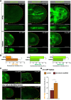
Ubx is required for expression of Timp in the haltere. (A) Maximum projection (Z-proj; top) and cross-sections (bottom) of 4 h wings and halteres expressing endogenous GFP-Timp, Mmp1-GFP or Mmp2-GFP. GFP-Timp is not detectable in the wing blade but is strongly expressed in specific regions of the haltere. Mmp2-GFP and Mmp1-GFP accumulate at the basal membrane of both the wing and the haltere. (B) Quantification of GFP-Timp, Mmp-GFP and Mmp2-GFP immunofluorescence signal in wings and halteres at 4 h APF. Halteres show higher levels of GFP-Timp and lower levels of Mmp1-GFP and Mmp2-GFP compared with the wing. (C) Maximum projections of GFP-Timp of control (ubx-Gal4/+) and ubxRNAi-expressing halteres (ubx-Gal4>UAS.UbxRNAi) at 4 h and 7 h APF. Depletion of Ubx in the haltere decreases GFP-Timp expression at 4 h APF, immediately before ectopic ECM degradation in mutant halteres. (D) Quantification of GFP-Timp immunofluorescence signal in control and ubx-Gal4>UAS.UbxRNAi wings and halteres at 4 h APF. Depletion of Ubx decreases GFP-Timp expression compared with control halteres. Data are mean±s.d., n>4 for each developmental stage. **P<0.005, ***P<0.001 (two-tailed Student's t-test). Dashed lines indicate the perimeter of the wing blade, determined by looking at the actin cytoskeleton. Scale bars: 50 μm.
Fig. 6.
Fig. 6.
Ubx binds to specific sites in the Timp, Mmp1 and Mmp2 genes in the haltere. (A) Ubx binding sites in the Timp gene from ChIP experiments performed in this study. To look for specific Ubx DNA binding sites in third instar larvae (L3) halteres, we extracted chromatin from L3 halteres and compared Ubx binding peaks in samples with and without adding the antibody to pulldown Ubx. We found two haltere-specific Ubx binding peaks for Timp (highlighted in red), located at 5′ and 3′ regulatory regions. (B) Ubx binding sites in the Mmp1 gene from ChIP experiments performed in this study. We found two haltere-specific Ubx binding peaks for Mmp1 (highlighted in red), located at 5′ intergenic regions or introns. The Mmp1-RF isoform that carries the GFP insertion in our Mmp1-GFP knock-in is marked with an asterisk. (C) Ubx binding sites in the Mmp2 gene from ChIP experiments performed in this study. We found two haltere-specific Ubx binding peaks for Mmp2 (highlighted in red), located at 5′ intergenic regions or introns. The Mmp2-RB isoform carrying the GFP insertion in our Mmp2-GFP knock-in is marked with an asterisk. Annotation of genomic location and protein isoforms were adapted from Flybase database (https://flybase.org/).
Fig. 7.
Fig. 7.
Preventing both basal and apical ECM remodelling strongly impairs wing morphogenesis. (A) Adult wings from control and animals overexpressing Timp (nub-Gal4>UAS.Timp), combined with the depletion of Sb (nub-Gal4>SbRNAi,UAS.Timp) or both Sb and Np (nub-Gal4>Np.RNAi;SbRNAi,UAS.Timp), compared with wings ectopically expressing a Ubx allele (nub-Gal4>UbxIa). Reduction in the activity or expression of both aECM and bECM proteases dramatically decreases wing size and length, resembling the wing-to-haltere transformation phenotype caused by UbxIa overexpression. (B) Maximum projections of 7 h APF wings from control and animals overexpressing Timp, combined with the depletion of Sb and Np, compared with wings ectopically expressing the UbxIa allele. Overexpression of Timp inhibits bECM degradation, whereas overexpression of Sb inhibits aECM degradation, impairing wing expansion and elongation (see Fig 1B), similar to UbxIa-expressing 7 h APF mutant wings. (C) Quantification of size (area) and shape characteristics (aspect ratio, dorsoventral adhesion and epithelial folding) in control (w), nub-Gal4>UAS.Timp, nub-Gal4>SbRNAi,UAS.Timp, nub-Gal4>Np.RNAi;SbRNAi,UAS.Timp and nub-Gal4>UbxIa wings compared with control (h) and Ubx-Gal4>Ubx.RNAi halteres. Inhibition of bECM degradation by Timp overexpression reduces wing size and dorsoventral adhesion. Data are mean±s.d. from up to 20 wings or halteres for each genotype. When combined with depletion of apical proteases, the defects associated with impaired bECM degradation in the wing increase and includes wing rounding, similar to wings ectopically expressing Ubx in the wing and control halteres. bECM depletion also results in folding of the wing blade. Scale bars: 50 μm.
Fig. 8.
Fig. 8.
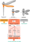
Ubx controls apical and basal ECM degradation to regulate morphogenesis. Schematic of Ubx expression and function in Drosophila and a hypothetical four-winged ancestor. Ubx controls organ shape via regulation of aECM and bECM proteases, in addition to its known functions in regulating organ growth and patterning. These target genes have presumably evolved to be specifically regulated in the Drosophila wing and/or haltere, and must be insensitive to Ubx in four-winged ancestors.

Comment in

Similar articles

Cited by

References

    1. Affolter M., Percival-Smith A., Muller M., Leupin W. and Gehring W. J. (1990a). DNA binding properties of the purified Antennapedia homeodomain. Proc. Natl. Acad. Sci. USA 87, 4093-4097. 10.1073/pnas.87.11.4093 - DOI - PMC - PubMed
    1. Affolter M., Schier A. and Gehring W. J. (1990b). Homeodomain proteins and the regulation of gene expression. Curr. Opin. Cell Biol. 2, 485-495. 10.1016/0955-0674(90)90132-X - DOI - PubMed
    1. Affolter M., Slattery M. and Mann R. S. (2008). A lexicon for homeodomain-DNA recognition. Cell 133, 1133-1135. 10.1016/j.cell.2008.06.008 - DOI - PubMed
    1. Agrawal P., Habib F., Yelagandula R. and Shashidhara L. S. (2011). Genome-level identification of targets of Hox protein Ultrabithorax in Drosophila: novel mechanisms for target selection. Sci. Rep. 1, 205 10.1038/srep00205 - DOI - PMC - PubMed
    1. Akam M. E. (1983). The location of Ultrabithorax transcripts in Drosophila tissue sections. EMBO J. 2, 2075-2084. 10.1002/j.1460-2075.1983.tb01703.x - DOI - PMC - PubMed

Publication types

MeSH terms