. 2020 Apr 3;295(14):4723-4732.
doi: 10.1074/jbc.RA119.011302. Epub 2020 Mar 2.

Mechanical stretch increases Kv1.5 current through an interaction between the S1-S2 linker and N-terminus of the channel

Affiliations

Mechanical stretch increases Kv1.5 current through an interaction between the S1-S2 linker and N-terminus of the channel

Alexandria O Milton et al. J Biol Chem. .

Abstract

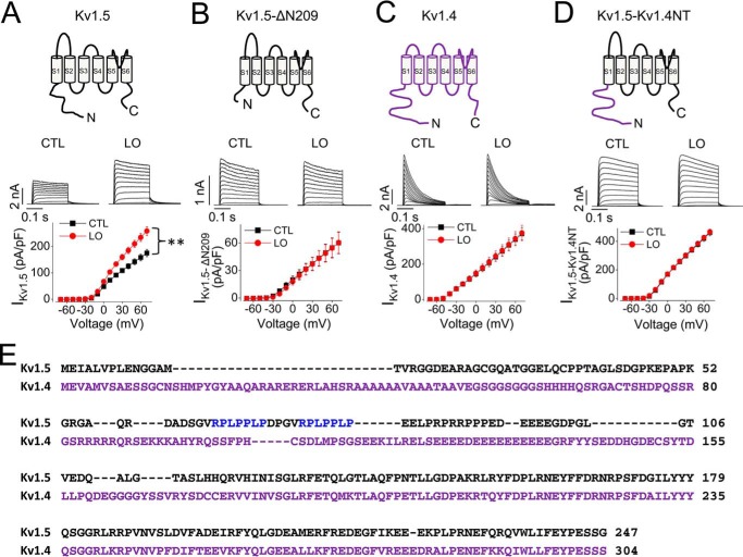
The voltage-gated potassium channel Kv1.5 plays important roles in atrial repolarization and regulation of vascular tone. In the present study, we investigated the effects of mechanical stretch on Kv1.5 channels. We induced mechanical stretch by centrifuging or culturing Kv1.5-expressing HEK 293 cells and neonatal rat ventricular myocytes in low osmolarity (LO) medium and then recorded Kv1.5 current (IKv1.5) in a normal, isotonic solution. We observed that mechanical stretch increased IKv1.5, and this increase required the intact, long, proline-rich extracellular S1-S2 linker of the Kv1.5 channel. The low osmolarity-induced IKv1.5 increase also required an intact intracellular N terminus, which contains the binding motif for endogenous Src tyrosine kinase that constitutively inhibits IKv1.5 Disrupting the Src-binding motif of Kv1.5 through N-terminal truncation or mutagenesis abolished the mechanical stretch-mediated increase in IKv1.5 Our results further showed that the extracellular S1-S2 linker of Kv1.5 communicates with the intracellular N terminus. Although the S1-S2 linker of WT Kv1.5 could be cleaved by extracellularly applied proteinase K (PK), an N-terminal truncation up to amino acid residue 209 altered the conformation of the S1-S2 linker and made it no longer susceptible to proteinase K-mediated cleavage. In summary, the findings of our study indicate that the S1-S2 linker of Kv1.5 represents a mechanosensor that regulates the activity of this channel. By targeting the S1-S2 linker, mechanical stretch may induce a change in the N-terminal conformation of Kv1.5 that relieves Src-mediated tonic channel inhibition and results in an increase in IKv1.5.

Keywords: Kv1.5; Src; Src kinase; cell biology; electrophysiology; ion channel; mechanical stretch; mechanotransduction; molecular biology; patch clamp; potassium channel; structure-function; voltage-gated potassium channel.


Conflict of interest statement

The authors declare that they have no conflicts of interest with the contents of this article

Figures

Figure 1.
Figure 1.
Centrifugation increases IKv1.5. Kv1.5-HEK cells were centrifuged (CENTR) at 70 × g for 5 min. Cells were then re-suspended in normal culture medium for 20 min prior to IKv1.5 recordings. Kv1.5-HEK cells without centrifugation were used as control (CTL). Representative current traces along with the voltage protocol (top) and summarized I-V relationship (bottom) are shown. CTL, n = 29; CENTR, n = 38; **, p < 0.01 at 20 mV and above.
Figure 2.
Figure 2.
LO medium treatment increases cell size and reversibly increases IKv1.5. A, culture of Kv1.5-HEK cells with LO medium for 30 min increased cell size (n = 13; **, p < 0.01). B, culture of Kv1.5-HEK cells with LO medium for 30 min increased IKv1.5. Representative current traces are depicted above the summarized I-V relationships (left) and activation curves (right). Activation curves were fitted to the Boltzmann function to determine V1/2 values and slope factors. CTL, n = 47; LO, n = 31; **, p < 0.01 at 20 mV and above for I-V curves; and tail currents at −30 mV following 50 mV depolarization for activation curves. C, LO-treatment mediated increase in IKv1.5 recovered (Recv) with time upon re-culture of cells in normal (isotonic) medium. Summarized I-V relationships at various time points were obtained from 7–27 cells. *, p < 0.05; **, p < 0.01 versus CTL at 20 mV and above; #, p < 0.05; ##, p < 0.01 versus LO at 20 mV and above.
Figure 3.
Figure 3.
LO medium treatment increases IKv1.5 in neonatal rat ventricular myocytes transfected with Kv1.5. Representative current traces are depicted above the summarized I-V relationship. CTL, n = 32; LO, n = 32. *, p < 0.05 at 20 mV and above.
Figure 4.
Figure 4.
Mechanical stretch induced by LO medium culture selectively increases IKv1.5. Currents from Kv1.5, Kv1.4, Kv4.3, Kv7.1+KCNE1, Kv10.1, and Kv11.1 channels were recorded from HEK 293 cells stably expressing the respective channels in control cells and cells treated with LO medium for 30 min. Representative current traces are depicted above the summarized I-V relationships. For IKv1.5, CTL, n = 47; LO, n = 31 (same set of data shown in Fig. 2B). For IKv1.4, CTL, n = 43; LO, n = 46; for IKv4.3, CTL, n = 42; LO, n = 42; for IKv7.1+KCNE1, CTL, n = 15; LO, n = 12; for IKv10.1, CTL, n = 18, LO, n = 18; for IKv11.1, CTL, n = 23, LO, n = 19. **, p < 0.01 at 20 mV and above.
Figure 5.
Figure 5.
The unique S1–S2 linker of Kv1.5 is involved in LO-mediated increase in IKv1.5. A, amino acid sequences of the S1–S2 linker of various Kv channels. Kv1.5 possesses an unusually long S1–S2 linker with 12 nonconserved proline residues (in magenta). The N-linked glycosylation site is shown in red. B, PK cleavage of the S1–S2 linker abolished LO-induced increase in IKv1.5. Schematic illustration of Kv1.5 PK cleavage (top) and representative current traces (middle) are depicted above the summarized I-V relationships (bottom). CTL, n = 14; LO, n = 21. C, mutating all 12 nonconserved prolines (P) to alanines (A) in the S1–S2 linker abolished LO-induced increase in IKv1.5. Schematic illustration of the Kv1.5–12PA mutant (top) as well as representative current traces (middle) are depicted above the summarized I-V relationships (bottom). CTL, n = 37; LO, n = 36. D, deletion of amino acid residues 282–300 in the S1–S2 linker abolished LO-induced increase in IKv1.5. Schematic illustration of Kv1.5-Δ282–300 mutant (top) as well as representative current traces (middle) are depicted above the summarized I-V relationships (bottom). CTL, n = 15, LO, n = 10. E, inhibition of glycosylation in the S1–S2 linker with tunicamycin (Tuni) treatment abolished the LO-induced increase in IKv1.5. Schematic illustration of WT Kv1.5 without glycosylation (top) as well as representative current traces (middle) are depicted above the summarized I-V relationships (bottom). CTL, n = 24; LO, n = 24.
Figure 6.
Figure 6.
The N terminus is involved in LO-mediated increase in IKv1.5. A, LO treatment for 30 min increased IKv1.5. Schematic illustration of WT Kv1.5 channel (top) and representative current traces (middle) are depicted above the summarized I-V relationships (bottom). CTL, n = 47; LO, n = 31; **, p < 0.01 at 20 mV and above (same set of data shown in Figs. 2B and 4 for Kv1.5). B, N terminus truncation mutant ΔN209 abolished LO-induced increase in IKv1.5. Schematic illustration of Kv1.5-ΔN209 mutant (top) and representative current traces (middle) are depicted above the summarized I-V relationships (bottom). CTL, n = 13; LO, n = 11. C, LO treatment for 30 min had no effects on IKv1.4. Schematic illustration of WT Kv1.4 channel (top) and representative current traces (middle) are depicted above the summarized I-V relationships (bottom). CTL, n = 43; LO, n = 46 (same set of data shown in Fig. 4 for Kv1.4); D, replacement of the N terminus of Kv1.5 with the N terminus of Kv1.4 prevented LO-induced increase in IKv1.5. Schematic illustration of Kv1.5–Kv1.4NT mutant (top) and representative current traces (bottom) are depicted above the summarized I-V relationships (bottom). CTL, n = 32; LO, n = 30. E, amino acid sequence alignment of the N termini of Kv1.5 (1–247, UniProt: P22460) and Kv1.4 (1–304, UniProt: P22459), with Kv1.5 SH3-binding motifs shown in blue.
Figure 7.
Figure 7.
Removal of Src-binding sites abolishes LO-mediated increase in IKv1.5. A, amino acid sequences showing the two consensus SH3–binding motifs (RPLPPLP, shown in blue) in the N terminus of Kv1.5 as well as the mutant Kv1.5-ΔPro, in which amino acids 64–82 were removed. B, effects of LO treatment on WT IKv1.5. CTL, n = 36; LO, n = 27; **, p < 0.01 at 20 mV and above. C, removal of the Src-binding sites in Kv1.5 prevented LO-induced increase in IKv1.5. Representative current traces (top) are depicted above the summarized I-V relationships (bottom). CTL, n = 26; LO, n = 28.
Figure 8.
Figure 8.
Effects of LO-treatment and Src inhibitor PP1 on Kv1.5 expression and function. A, LO treatment did not affect the total amount of Kv1.5 proteins but increased the cell surface mature channel expression. The density of the 75-kDa band in LO-treated cells was normalized to that of control cells in the same gel and shown in the scatter plots (n = 5). B, PP1 treatment did not affect the total amount of Kv1.5 proteins but increased the cell surface mature channel expression. The density of the 75-kDa band in PP1-treated cells was normalized to that of control cells in the same gel and shown in the scatter plot (n = 5). For A and B, boxes represent interquartile ranges, horizontal lines represent medians, whiskers represent 5–95% ranges, and gray boxes represent means. **, p < 0.01 versus CTL. C, Src inhibitor PP1 treatment increased IKv1.5 and prevented LO-mediated increase in IKv1.5. Representative current traces (top) are depicted above the summarized I-V relationships (bottom). CTL, n = 32; PP1, n = 35; LO, n = 26; PP1+LO, n = 28. *, p < 0.05 at 20 mV and above, compared with control (CTL). There was no significant difference among PP1, LO, and PP1+LO groups.
Figure 9.
Figure 9.
The Kv1.5 S1–S2 linker communicates with the N terminus in a conformational manner. Truncation of N terminus altered the susceptibility of the S1–S2 linker to PK cleavage. WT Kv1.5 displays 75-kDa and 68-kDa bands on Western blot analysis. The 75-kDa band represents the mature, fully glycosylated channel protein in the plasma membrane, whereas the 68-kDa band represents the immature channel protein inside the cell. ΔN209 Kv1.5 also presents as two bands; the 50-kDa band represents mature protein in the plasma membrane, the 43-kDa band represents immature protein inside the cell. Although PK completely cleaved the mature (cell surface) channel proteins of WT Kv1.5, it failed to cleave the mature (cell surface) channel proteins of ΔN209 Kv1.5 (n = 6).

Similar articles

Cited by

References

    1. Fedida D., Wible B., Wang Z., Fermini B., Faust F., Nattel S., and Brown A. M. (1993) Identity of a novel delayed rectifier current from human heart with a cloned K+ channel current. Circ. Res. 73, 210–216 10.1161/01.RES.73.1.210 - DOI - PubMed
    1. Wang Z., Fermini B., and Nattel S. (1993) Sustained depolarization-induced outward current in human atrial myocytes: Evidence for a novel delayed rectifier K+ current similar to Kv1.5 cloned channel currents. Circ. Res. 73, 1061–1076 10.1161/01.RES.73.6.1061 - DOI - PubMed
    1. Archer S. L., Souil E., Dinh-Xuan A. T., Schremmer B., Mercier J. C., El Yaagoubi A., Nguyen-Huu L., Reeve H. L., and Hampl V. (1998) Molecular identification of the role of voltage-gated K+ channels, Kv1.5 and Kv2.1, in hypoxic pulmonary vasoconstriction and control of resting membrane potential in rat pulmonary artery myocytes. J. Clin. Invest. 101, 2319–2330 10.1172/JCI333 - DOI - PMC - PubMed
    1. Ohanyan V., Yin L., Bardakjian R., Kolz C., Enrick M., Hakobyan T., Kmetz J., Bratz I., Luli J., Nagane M., Khan N., Hou H., Kuppusamy P., Graham J., Fu F. K., et al. (2015) Requisite role of Kv1.5 channels in coronary metabolic dilation. Circ. Res. 117, 612–621 10.1161/CIRCRESAHA.115.306642 - DOI - PMC - PubMed
    1. Hogan-Cann A., Li W., Guo J., Yang T., and Zhang S. (2016) Proteolytic cleavage in the S1-S2 linker of the Kv1.5 channel does not affect channel function. Biochim. Biophys. Acta 1858, 1082–1090 10.1016/j.bbamem.2016.02.012 - DOI - PubMed

Publication types

MeSH terms

Substances