. 2020 Mar 17;117(11):6121-6128.
doi: 10.1073/pnas.1917748117. Epub 2020 Mar 2.

Cell-penetrating peptide inhibits retromer-mediated human papillomavirus trafficking during virus entry

Affiliations

Cell-penetrating peptide inhibits retromer-mediated human papillomavirus trafficking during virus entry

Pengwei Zhang et al. Proc Natl Acad Sci U S A. .

Abstract

Virus replication requires critical interactions between viral proteins and cellular proteins that mediate many aspects of infection, including the transport of viral genomes to the site of replication. In human papillomavirus (HPV) infection, the cellular protein complex known as retromer binds to the L2 capsid protein and sorts incoming virions into the retrograde transport pathway for trafficking to the nucleus. Here, we show that short synthetic peptides containing the HPV16 L2 retromer-binding site and a cell-penetrating sequence enter cells, sequester retromer from the incoming HPV pseudovirus, and inhibit HPV exit from the endosome, resulting in loss of viral components from cells and in a profound, dose-dependent block to infection. The peptide also inhibits cervicovaginal HPV16 pseudovirus infection in a mouse model. These results confirm the retromer-mediated model of retrograde HPV entry and validate intracellular virus trafficking as an antiviral target. More generally, inhibiting virus replication with agents that can enter cells and disrupt essential protein-protein interactions may be applicable in broad outline to many viruses.

Keywords: HPV; antiviral agent; protein transduction domain; retrograde; retromer.


Conflict of interest statement

Competing interest statement: P.Z. and D.D. are inventors on a patent application related to this work.

Figures

Fig. 1.
Fig. 1.
Identification and analysis of peptides that inhibit HPV infection. (A, Top) Sequences of L2 peptides with wild-type CPP segments (red) and RBS (green) highlighted. Amino acid substitutions in the CPP or RBS mutant peptides are shown in blue. (A, Bottom) RBS and flanking sequences of indicated HPV L2 proteins or DMT1-II. (B) Inhibitory dose–response curve for the wild-type P16/16 peptide. HeLa cells were pretreated with various concentrations of P16/16 for 1 h prior to infection with HPV16 PsV at an MOI of 1. The peptide and PsV were left in the medium for the duration of the experiment. At 48 h.p.i., flow cytometry was used to determine the fraction of cells expressing reporter protein HcRed. The graph shows mean results of three experiments, +/− SD. (C) HeLa cells were pretreated for 1 h with 14 µM (orange line) or 28 µM (green line) P16/16 or 14 µM PDM/16 (gray line), or left untreated (blue line). Cells were then infected with HPV16 PsV at the indicated MOI, and at 48 h.p.i. infectivity was measured by flow cytometry for reporter gene expression as in B. (D) Inhibition of authentic HPV16. HeLa cells were infected with HPV16 harvested from organotypic cultures of human keratinocytes or with HPV16 PsV in the presence (gray bars) or absence (black bars) of 14 μM P16/16. Infection by HPV16 and HPV16 PsV was assessed by qRT-PCR for expression of HPV E7 and HcRed mRNA, respectively, and normalized to infection by the cognate virus in the absence of the peptide. The graph shows average results of three independent experiments, +/− SD, where infection of untreated cells is set at 100%. The background signal determined with noncognate primers was <0.01%. (E) Inhibition of HPV infection of HaCaT cells. HaCaT keratinocytes were infected at an MOI of 1 with HPV16 PsV in the presence (gray bars) or absence (black bars) of 14 μM P16/16, and infectivity was assessed at 48 h.p.i. by flow cytometry for HcRed fluorescence and displayed as in B. (F) HeLa cells were treated for 24 h with no peptide (black bars) or 14 µM P16/16 (gray bars). The peptide was then removed, and the cells were incubated for the indicated period of time prior to infection with HPV16 PsV at an MOI of 1. At 48 h.p.i, infectivity was measured by flow cytometry as in Fig. 1B. The graph shows results of three experiments +/− SD, normalized to no peptide control at time = 0. Numbers indicate the P value for each pairwise comparison.
Fig. 2.
Fig. 2.
Identification of sequence elements required for inhibition of multiple HPV types. HeLa cells were pretreated with 14 µM indicated peptide for 1 h, followed by infection with HPV16 (blue), HPV18 (green), or HPV5 PsV (orange) at an MOI of 1. Peptides and PsV were left in the medium for the duration of the experiment. As a control, cells were incubated with the solution used to dissolve the peptide. At 48 h.p.i., flow cytometry was used to determine the fraction of cells expressing HcRed. The graph shows average results of three independent experiments, +/− SD, normalized to infection of each PsV type in the absence of the peptide. P16/16, P16/Tat, and P16/31 caused statistically significant (P < 0.01 or lower) inhibition of all three PsV types, which was blocked by the mutations. P values are shown for comparison of P16/16 and no peptide, P16/6A, P16/3R, and PDM/16 for HPV16 PsV. Similar levels of significance were achieved with these peptides and HPV18 and HPV5 PsV.
Fig. 3.
Fig. 3.
The peptide binds retromer and blocks retromer association with HPV. (A) Uninfected HeLa cells were incubated with 14 μM bP16/16 or bPDM/16 for 3 h. Cells were then fixed, permeabilized, and stained with Alexa Fluor streptavidin (green) and an antibody recognizing the retromer subunit, VPS35 (red). The overlapping signal is pseudocolored yellow in the merged panels. Nuclei were stained blue with DAPI. Cells were imaged by confocal microscopy. (B) Mander’s correlation coefficients for overlap between streptavidin and VPS35, with each spot representing the data for an individual cell. Approximately 100 cells were analyzed for each condition. Each dot represents data from a single cell. Horizontal lines show mean and SD. (C) HeLa cells were treated for 6 h with bP16/16. Medium was replaced with fresh medium without the peptide and the cells were incubated at 37 °C for the indicated period of time before staining with fluorescent streptavidin as in A. (D) HeLa cells were incubated for 1 h without the peptide or with 14 μM P16/16 or PDM/16, followed by mock-infection or infection with HPV16 PsV at an MOI of 200. At 8 h.p.i., cells were fixed and processed for PLA with anti-L1 and anti-VPS35 antibodies. The PLA signal is green, and the nuclei are stained blue with DAPI. Cells were imaged by confocal microscopy. (E) Approximately 200 cells in each sample were imaged. Images were processed by Fiji software and analyzed by BlobFinder software to measure total fluorescence intensity per cell in each sample. The graph shows the mean fluorescence per cell and SD, normalized to that of cells infected with wild-type HPV16 PsV in the absence of the peptide from three independent experiments (N, HPV no peptide; W, HPV + P16/16; M, HPV + PDM/16; n.s., not significant).
Fig. 4.
Fig. 4.
The peptide inhibits HPV exit from the endosome. HeLa cells were incubated for 1 h with or without 14 μM P16/16, followed by mock-infection or infection with HPV16 PsV at an MOI of 200. At 8 and 16 h.p.i., PLA was performed as described in Fig. 3D with anti-L1 and anti-EEA1 antibody (A) or TGN46 antibody (B). The PLA signal for EEA1-L1 was normalized to that of cells infected with HPV16 PsV in the absence of peptide at 8 h.p.i., and the TGN46-L1 signal was normalized to untreated cells at 16 h.p.i. The graphs show average normalized fluorescence per cell and SD for three independent experiments (n.s., not significant). (C, Left) HeLa cells were mock-infected or infected with HPV16 PsV containing EdU-labeled reporter plasmid DNA at an MOI of 50. Where indicated, cells were pretreated for 1 h with 14 μM P16/16. At 48 h.p.i., cells were fixed and treated with Click-iT chemistry to stain viral DNA (green) and incubated with anti-PML antibody (red). The overlap in EdU and PML staining is pseudocolored yellow. Nuclei are stained blue. (C, Right) Nuclear and nonnuclear EdU staining as in C, Left, was quantified for 60 cells in each condition. Each dot represents data from an individual cell. Horizontal lines indicate mean and SD.
Fig. 5.
Fig. 5.
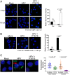
Fate of HPV following peptide inhibition. (A) HeLa cells were incubated for 1 h with or without 14μM P16/16, followed by mock-infection or infection with HPV16 PsV containing FLAG-tagged L2 at an MOI of 50. At the indicated times after infection, cells were permeabilized and stained with anti-FLAG (red) and anti-L1 (green) antibody. Nuclei are stained blue. The overlap in FLAG and L1 staining is pseudocolored yellow in the merged panels. There was no antibody staining in mock-infected cells. (B) Cells pretreated with 14 µM P16/16 (+) or left untreated (−) were mock-infected or infected with HPV16 PsV at an MOI of 20 as in A. At the indicated h.p.i., extracts were prepared and assessed by immunoblotting for L1 and L2 expression. GADPH acts as a loading control. (C) Cells treated with P16/16 or left untreated were infected as in A. Where indicated, cells were treated with 100 µM chloroquine starting 30 min prior to infection. At 16 h.p.i., cells were permeabilized and stained with anti-FLAG (green) and anti-LAMP1 (red) antibodies. Nuclei are stained blue. The overlap between FLAG and LAMP1 staining is pseudocolored yellow. (D) Overlapping staining as in C was quantified as in Fig. 3B. (E) Cells were infected and treated with p16/16 and chloroquine (CQ) as in C. Extracts were prepared at 16 h.p.i and analyzed by immunoblotting.
Fig. 6.
Fig. 6.
The peptide inhibits HPV pseudovirus infection in vivo. Female FVB mice were treated with Depo-Provera and Conceptrol as described in Materials and Methods. Mice were then treated intravaginally with 4% carboxymethyl cellulose (CMC) (designated mock) or 4% CMC containing 3 × 10⁵ transducing units of HPV16:pCLucF plus or minus 25 μg of P16/16 or PDM/16. Two days later, reproductive tract tissues were harvested and assayed in triplicate in vitro for luciferase activity, which is reported as relative light units per μg total protein. Each dot represents data from an individual mouse. Statistical analyses were made with the two-sided Wilcoxon Rank Sum test.

Similar articles

Cited by

References

    1. Buck C. B., et al. , Arrangement of L2 within the papillomavirus capsid. J. Virol. 82, 5190–5197 (2008). - PMC - PubMed
    1. Campos S. K., Subcellular trafficking of the papillomavirus genome during initial infection: The remarkable abilities of minor capsid protein L2. Viruses 9, E370 (2017). - PMC - PubMed
    1. Zhang P., Monteiro da Silva G., Deatherage C., Burd C., DiMaio D., Cell-penetrating peptide mediates intracellular membrane passage of human papillomavirus L2 protein to trigger retrograde trafficking. Cell 174, 1465–1476.e13 (2018). - PMC - PubMed
    1. Burd C., Cullen P. J., Retromer: A master conductor of endosome sorting. Cold Spring Harb. Perspect. Biol. 6, a016774 (2014). - PMC - PubMed
    1. Day P. M., Thompson C. D., Schowalter R. M., Lowy D. R., Schiller J. T., Identification of a role for the trans-Golgi network in human papillomavirus 16 pseudovirus infection. J. Virol. 87, 3862–3870 (2013). - PMC - PubMed

Publication types

MeSH terms

Substances

LinkOut - more resources