. 2020 Mar 17;117(11):5791-5800.
doi: 10.1073/pnas.1920251117. Epub 2020 Mar 2.

bioPROTACs as versatile modulators of intracellular therapeutic targets including proliferating cell nuclear antigen (PCNA)

Affiliations

bioPROTACs as versatile modulators of intracellular therapeutic targets including proliferating cell nuclear antigen (PCNA)

Shuhui Lim et al. Proc Natl Acad Sci U S A. .

Abstract

Targeted degradation approaches such as proteolysis targeting chimeras (PROTACs) offer new ways to address disease through tackling challenging targets and with greater potency, efficacy, and specificity over traditional approaches. However, identification of high-affinity ligands to serve as PROTAC starting points remains challenging. As a complementary approach, we describe a class of molecules termed biological PROTACs (bioPROTACs)-engineered intracellular proteins consisting of a target-binding domain directly fused to an E3 ubiquitin ligase. Using GFP-tagged proteins as model substrates, we show that there is considerable flexibility in both the choice of substrate binders (binding positions, scaffold-class) and the E3 ligases. We then identified a highly effective bioPROTAC against an oncology target, proliferating cell nuclear antigen (PCNA) to elicit rapid and robust PCNA degradation and associated effects on DNA synthesis and cell cycle progression. Overall, bioPROTACs are powerful tools for interrogating degradation approaches, target biology, and potentially for making therapeutic impacts.

Keywords: PCNA; bioPROTAC; targeted degradation.


Conflict of interest statement

The authors declare no competing interest.

Figures
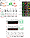
Fig. 1.
Fig. 1.
vhhGFP4-SPOP induces the ubiquitin-mediated proteasomal degradation of H2B-GFP. (A) Design of the chimeric protein vhhGFP4-SPOP167–374 for the degradation of GFP-tagged proteins. The substrate-binding MATH domain of the E3 adaptor SPOP (amino acids 1–166) was replaced by vhhGFP4 (25, 26), a single-domain antibody fragment that binds GFP. This will enable the ubiquitin tagging of GFP fusion proteins such as H2B-GFP by the CUL3-based CRL complex. Protein Data Bank (PDB) structures are shown for SPOP (3HQI) and GFP:GFP-nanobody complex (3OGO). (B) Flow cytometric analysis of H2B-GFP/HEK293 Tet-On 3G cells transiently transfected with various bidirectional, Tet-responsive plasmids. Doxycycline (100 ng/mL) was added to induce the simultaneous expression of mCherry and vhhGFP4-SPOP167–374 (or its controls). vhhGFP4mut lacks the complementarity determining region 3 (CDR3) and cannot bind GFP. SPOPmut lacks the three-box motif and cannot bind CUL3. GFP and mCherry fluorescence intensities were measured 24 h after doxycycline induction. (C) Confocal imaging analysis of the same set of cells as in B. Plasma membrane (pseudocolored white) was labeled using the CellMask Deep Red plasma membrane stain. Yellow arrow denotes an example of a transfected cell (mCherry positive) that have lost H2B-GFP. (D) Western blot analysis of H2B-GFP/HEK293 Tet-On 3G cells treated as in B and sorted according to the levels of mCherry using FACS. Gating was set such that mCherry (−) cells have the same signal intensities as untreated cells in the mCherry channel, and anything above this basal level was assigned mCherry (+). Expression of vhhGFP4-SPOP167–374 (or its controls) was detected using an anti-FLAG-tag antibody (Left Lower, red bands, n = 3 for vhhGFP4-SPOP167–374). The substrate H2B-GFP was detected using an anti-GFP antibody (Left Lower row, green bands), and band intensities were quantified and normalized to the levels of the loading control Hsp90 (Right). (E) Western blot analysis of H2B-GFP/HEK293 Tet-On 3G cells transiently transfected with the mCherry/vhhGFP4-SPOP167–374 bidirectional inducible plasmid and treated with the indicated concentrations of doxycycline and MG132 (a proteasome inhibitor) for 16 h. FACS sorting was conducted as in D. Band intensities of H2B-GFP and FLAG-tagged vhhGFP4-SPOP167–374 were quantified and plotted in Right. See SI Appendix, Fig. S5 for uncropped blots and expected molecular weight of each protein. (F) Flow cytometric analysis of the same set of cells as in E.
Fig. 2.
Fig. 2.
Flexibility in the type of binder used for generating bioPROTACs. Flow cytometric analysis of H2B-GFP/HEK293 Tet-On 3G cells transiently transfected with various bidirectional, Tet-responsive plasmids. Doxycycline (100 ng/mL) was added for 24 h to induce the simultaneous expression of mCherry and the different GFP binders fused to SPOP167–374. A total of seven GFP binders were tested: one nanobody (vhhGFP4; refs. and 26), one DARPin (3G86.32; ref. 29), two αReps (bGFP-A and bGFP-C; ref. 30), and three monobodies (GS2, GL6, and GL8; ref. 31). The values (in green) on the scatter plots indicate the percentage of GFP-negative cells in the mCherry-positive transfected population, which corresponds to successful H2B-GFP depletion by the respective SPOP-based anti-GFP bioPROTAC. The table summarizes the molecular mass of the GFP binders and their reported binding affinities to GFP. Representative PDB structures are shown for each scaffold (3OGO, 2QYJ, 4XVP, 1TTG), alpha helixes are colored blue, and beta strands are colored red.
Fig. 3.
Fig. 3.
Flexibility in the type of E3 ubiquitin ligase used for generating bioPROTACs. (A) Table of 10 different substrate recognition subunit (SRS) used in bioPROTAC designs to explore alternative E3 ligases. Information regarding each SRS are listed as follows: the E3 ubiquitin ligase complex they function in, PDB structural information, National Center for Biotechnology Information protein accession number, region of the protein fused to vhhGFP4, molecular mass of the region fused to vhhGFP4 and previous records of their use in PROTAC strategies. Truncations were designed to replace the original substrate-binding domain with the GFP-binding nanobody vhhGFP4. (B) Flow cytometric analysis of H2B-GFP/HEK293 Tet-On 3G cells transiently transfected with various bidirectional, Tet-responsive plasmids. Doxycycline (100 ng/mL) was added for 24 h to induce the simultaneous expression of mCherry and the different truncated SRSs fused to vhhGFP4. A total of 10 truncated SRSs was tested, grouped according to the cullin E3 scaffold they recruit. The values (in green) on the scatter plots indicate the percentage of GFP-negative cells in the mCherry-positive transfected population, which corresponds to successful H2B-GFP depletion by the respective vhhGFP4-based anti-GFP bioPROTAC. PDB structures are shown for each SRS, alpha helixes are colored blue, and beta strands are colored red. The dotted area represents the portion fused to vhhGFP4.
Fig. 4.
Fig. 4.
bioPROTACs inform on substrate degradability and E3 selection. Flow cytometric analysis of HEK293 Tet-On 3G cells with stable integration of GFP, H2B-GFP, or PCNA-GFP. Each of the three stable cell lines was transiently transfected with the same panel of bidirectional, Tet-responsive plasmids. Doxycycline (100 ng/mL) was added for 24 h to induce the simultaneous expression of mCherry and the different anti-GFP bioPROTAC. Cells in Q1 represent successful H2B-GFP depletion by the respective anti-GFP bioPROTAC. (Bottom) Confocal imaging analysis of PCNA-GFP/HEK293 Tet-On 3G cells transiently transfected with various bidirectional, Tet-responsive plasmids. Doxycycline (100 ng/mL) was added for 24 h to induce the simultaneous expression of mCherry and the different anti-GFP bioPROTACs.
Fig. 5.
Fig. 5.
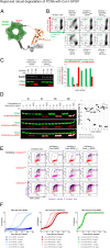
Rapid and robust degradation of PCNA with Con1-SPOP. (A) Design of the chimeric protein Con1-SPOP167–374 for the degradation of PCNA. The substrate-binding MATH domain of the E3 adaptor SPOP (amino acids 1–166) was replaced by Con1, a high-affinity peptide ligand of PCNA. This will enable the ubiquitin tagging of PCNA by the CUL3-based CRL complex. PDB structures are shown for SPOP (3HQI) and PCNA (1AXC). (B) Flow cytometric analysis of PCNA-GFP/HEK293 Tet-On 3G cells transiently transfected with various bidirectional, Tet-responsive plasmids. Doxycycline (100 ng/mL) was added for 24 h to induce the simultaneous expression of mCherry and the different chimeric proteins. vhhGFP4mut lacks the complementarity determining region 3 (CDR3) and cannot bind GFP. SPOPmut lacks the three-box motif and cannot bind CUL3. Con1mut bears point mutations in the three critical PCNA-interacting residues and cannot bind PCNA. The values (in green) on the scatter plots indicate the percentage of GFP-negative cells in the mCherry-positive transfected population, which corresponds to successful PCNA-GFP depletion by the respective SPOP-based bioPROTAC. (C) Western blot analysis of HEK293 Tet-On 3G cells transiently transfected and induced with doxycycline as in B and sorted according to the levels of mCherry using FACS. Gating was set such that mCherrylow cells have the same signal intensities as untreated cells in the mCherry channel, whereas mCherrymid and mCherryhigh cells have increasing levels of mCherry fluorescence. Expression of Con1-SPOP167–374 (or its control) was detected using an anti-FLAG-tag antibody (Left Lower, red bands) and the expected molecular mass of each chimeric protein is indicated in kilodaltons. The substrate PCNA was detected using an anti-PCNA antibody (Left Lower, green bands). Band intensities of FLAG-tagged vhhGFP4-SPOP167–374/SPOPmut and endogenous PCNA were quantified and normalized to the levels of the loading control Hsp90 (Right). (D) Western blot analysis of T-REx-293 cells with stable integration of Con1-SPOP167–374 (or its controls) under the control of a Tet-responsive promoter. Various concentrations of doxycycline were added to the culture media for the indicated length of time and lysates were collected. Proteins were detected as in C. PCNA levels were quantified and expressed as fold of untreated cells. (E) EdU labeling and flow cytometric analysis of HEK293 Tet-On 3G cells transiently transfected and induced with doxycycline as in B. Cells undergoing DNA synthesis were labeled with 10 µM EdU for 2 h and the percentage of EdU-positive S-phase cells (in purple) was expressed according to the level of mCherry signal intensities. Gating for mCherry expression was performed as in C. (F) Incucyte confluency measurements of T-REx-293 cells with stable integration of Con1-SPOP167–374 (or its controls) under the control of a Tet-responsive promoter. Various concentrations of doxycycline were added to the culture media and the percentage confluency of the cells was tracked continuously over 10 d.
Fig. 6.
Fig. 6.
Summary of the different modalities that can be used for the inhibition of a POI. The availability of ligands, the mode of inhibition (stoichiometric versus degradation), and the ease of delivery are parameters that can influence the tractability of each approach. By working at the protein level, bioPROTACs reduces risks associated with genetic manipulation using CRISPR and off-target effects commonly seen with siRNA. Compared to small molecules and peptides, ligands used in bioPROTAC approaches are easier to discover. However, delivery challenges need to be addressed and options include the delivery of bioPROTAC mRNA (refer to Discussion).

References

    1. Toure M., Crews C. M., Small-molecule PROTACS: New approaches to protein degradation. Angew. Chem. Int. Ed. Engl. 55, 1966–1973 (2016). - PubMed
    1. Gechijian L. N., et al. , Functional TRIM24 degrader via conjugation of ineffectual bromodomain and VHL ligands. Nat. Chem. Biol. 14, 405–412 (2018). - PMC - PubMed
    1. Huang X., Dixit V. M., Drugging the undruggables: Exploring the ubiquitin system for drug development. Cell Res. 26, 484–498 (2016). - PMC - PubMed
    1. Churcher I., Protac-induced protein degradation in drug discovery: Breaking the rules or just making new ones? J. Med. Chem. 61, 444–452 (2018). - PubMed
    1. Burslem G. M., et al. , The Advantages of targeted protein degradation over inhibition: An RTK case study. Cell Chem. Biol. 25, 67–77.e3 (2018). - PMC - PubMed

MeSH terms

Substances