. 2020 Mar 17;117(11):6047-6055.
doi: 10.1073/pnas.1920413117. Epub 2020 Mar 2.

Lactate dehydrogenase inhibition synergizes with IL-21 to promote CD8+ T cell stemness and antitumor immunity

Affiliations

Lactate dehydrogenase inhibition synergizes with IL-21 to promote CD8+ T cell stemness and antitumor immunity

Dalton Hermans et al. Proc Natl Acad Sci U S A. .

Abstract

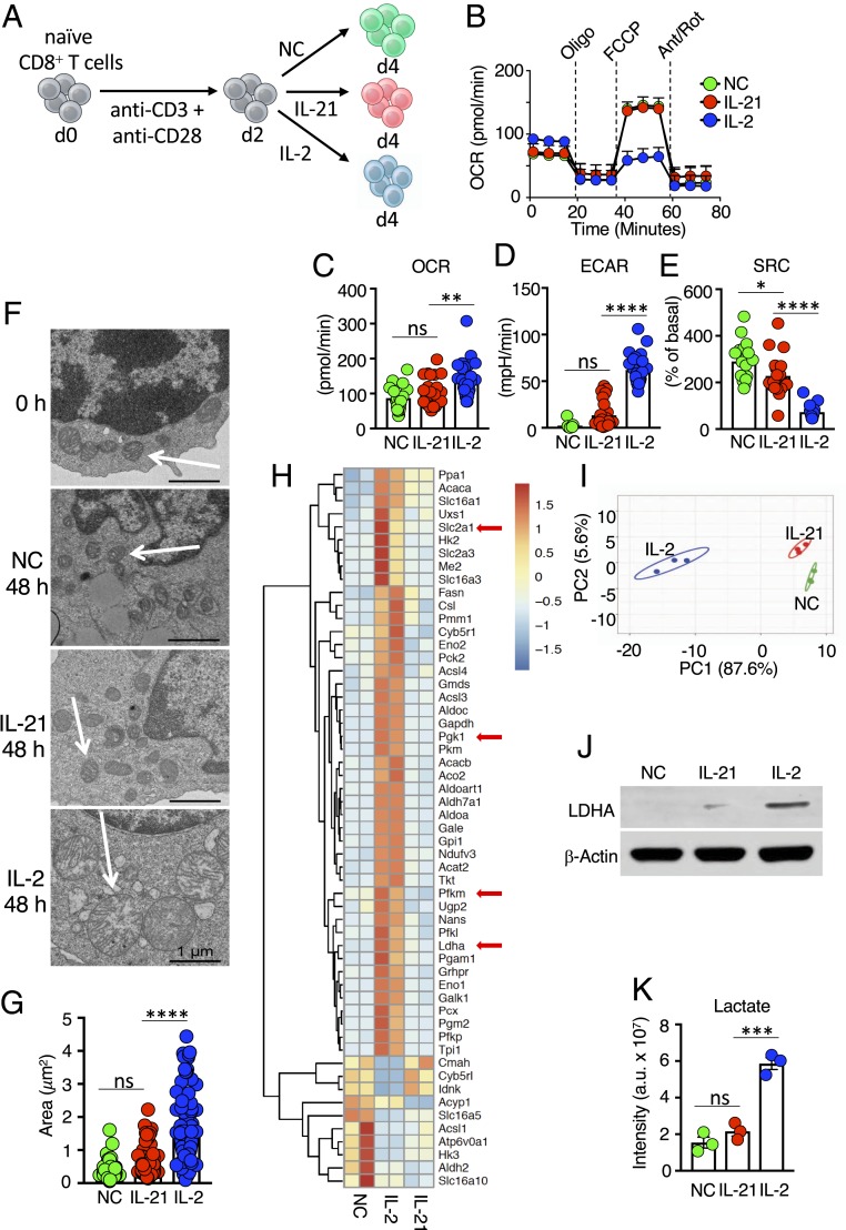
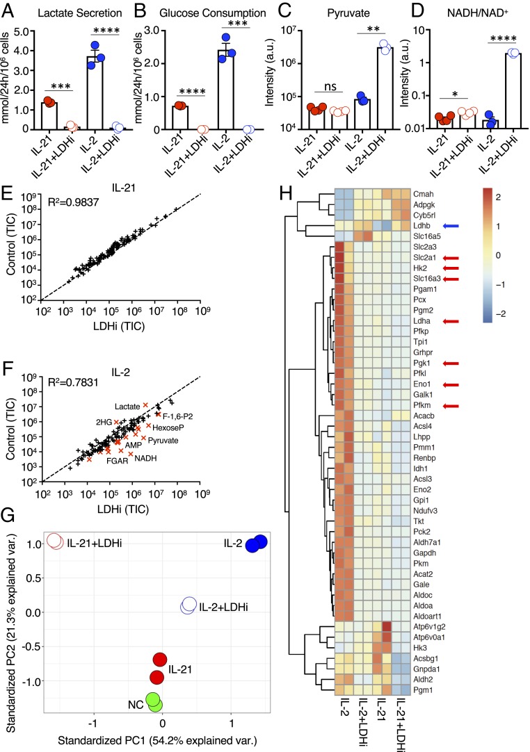
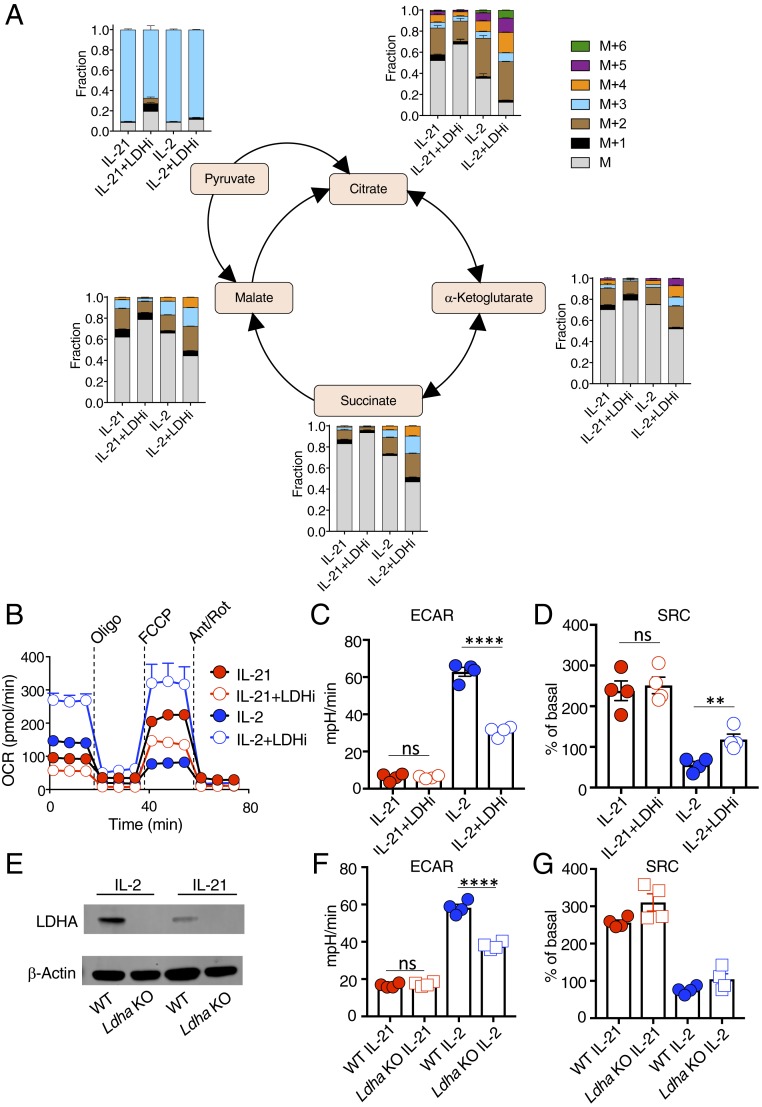
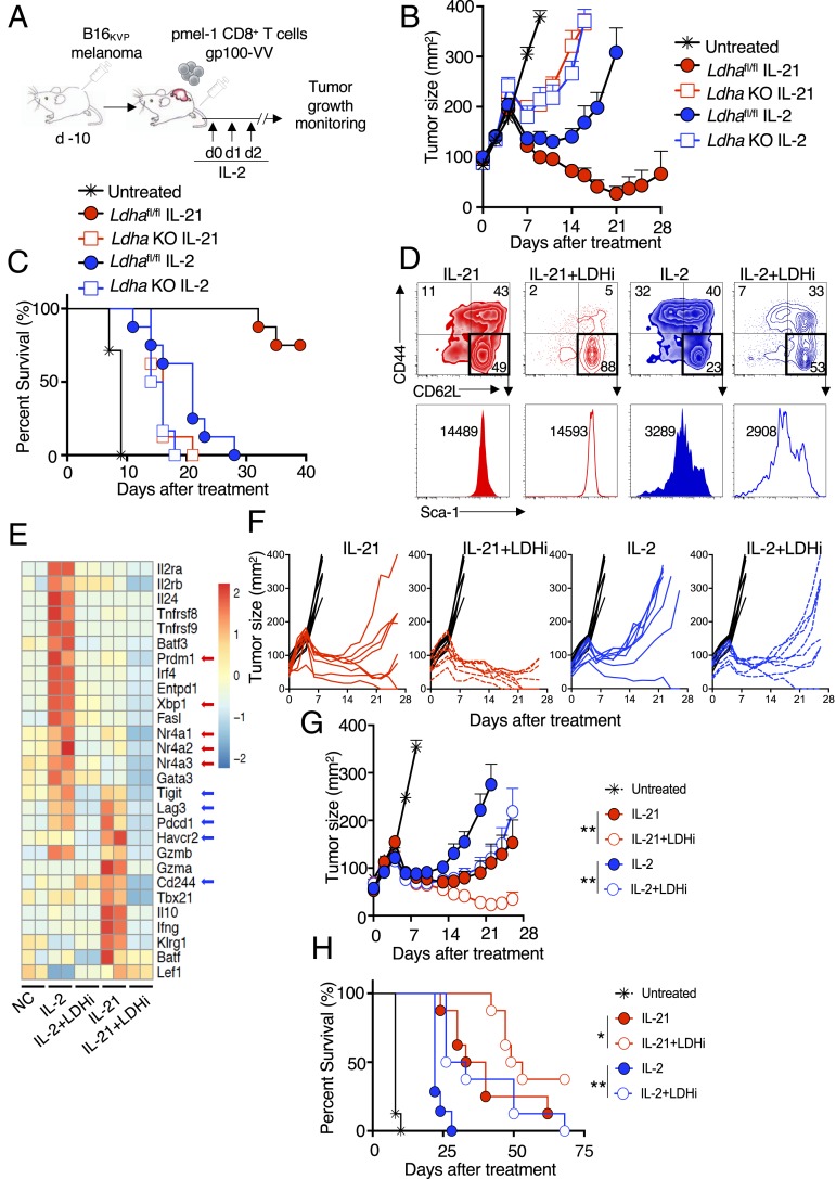
Interleukin (IL)-2 and IL-21 dichotomously shape CD8+ T cell differentiation. IL-2 drives terminal differentiation, generating cells that are poorly effective against tumors, whereas IL-21 promotes stem cell memory T cells (TSCM) and antitumor responses. Here we investigated the role of metabolic programming in the developmental differences induced by these cytokines. IL-2 promoted effector-like metabolism and aerobic glycolysis, robustly inducing lactate dehydrogenase (LDH) and lactate production, whereas IL-21 maintained a metabolically quiescent state dependent on oxidative phosphorylation. LDH inhibition rewired IL-2-induced effects, promoting pyruvate entry into the tricarboxylic acid cycle and inhibiting terminal effector and exhaustion programs, including mRNA expression of members of the NR4A family of nuclear receptors, as well as Prdm1 and Xbp1 While deletion of Ldha prevented development of cells with antitumor effector function, transient LDH inhibition enhanced the generation of memory cells capable of triggering robust antitumor responses after adoptive transfer. LDH inhibition did not significantly affect IL-21-induced metabolism but caused major transcriptomic changes, including the suppression of IL-21-induced exhaustion markers LAG3, PD1, 2B4, and TIM3. LDH inhibition combined with IL-21 increased the formation of TSCM cells, resulting in more profound antitumor responses and prolonged host survival. These findings indicate a pivotal role for LDH in modulating cytokine-mediated T cell differentiation and underscore the therapeutic potential of transiently inhibiting LDH during adoptive T cell-based immunotherapy, with an unanticipated cooperative antitumor effect of LDH inhibition and IL-21.

Keywords: IL-2; IL-21; LDH; adoptive immunotherapy; immunometabolism.


Conflict of interest statement

Competing interest statement: W.J.L. is an inventor on patents or patent applications related to IL-2 and IL-21. D.H., S.G., L.M.N., L.G., and W.J.L. are inventors on patent application(s) related to the LDH inhibitor described herein. L.G. is an inventor on a patent related to methods for generating TSCM cells.

Figures

Fig. 1.
Fig. 1.
Distinctive metabolic effects of IL-2 versus IL-21. (A) Schematic of protocol for stimulating CD8+ T cells. Naive T cells were isolated, activated with anti-CD3 and anti-CD28 for 2 d, and cultured with NC, IL-21, or IL-2 for another 2 d. (B) Seahorse experiment measuring the OCR for CD8+ T cells treated with NC, IL-21, or IL-2 following treatment with oligomycin, FCCP, and antimycin A/rotenone. Data are representative of 11 independent experiments. (CE) Bar graphs indicating basal OCR (C), ECAR (D), and SRC (E), as measured from Seahorse assays. (F) Electron micrographs are representative images from two independent experiments. (Scale bar: 1 μm.) (G) Calculated area of mitochondria from electron micrographs from cells treated with IL-2, IL-21, or NC. (H) RNA-Seq heatmap of differentially expressed metabolic genes from cells treated with NC, IL-2, or IL-21. Shown is the scale for fold induction or repression. (I) Principal component analysis plot based on metabolomics data of cells treated with NC, IL-2, or IL-21. (J) Western blotting of LDHA protein. (K) LC-MS–based analysis of intracellular lactate. *P < 0.05; **P < 0.01; ***P < 0.001; ****P < 0.0001; ns, not significant.
Fig. 2.
Fig. 2.
LDH inhibition alters glycolytic flux and transcriptional programming. (A and B) Using a YSI 2900 Series Biochemical Analyzer, media measurements for lactate secretion (A), and glucose consumption (B) were determined for cells treated with IL-2 or IL-21 without or with LDHi. (C and D) Pyruvate levels (C) and intracellular NADH/NAD+ ratio (D) were assessed by LC-MS for cells treated with IL-2 or IL-21 in the absence or presence of LDHi. (E and F) Truth plot showing correlation of intracellular metabolites for cells treated with IL-21 versus those treated with IL-21+LDHi (E) or IL-2 versus IL-2+LDHi (F); in F, the metabolites in red had a fold change >4. (G) Principal component analysis generated from RNA-Seq data of cells treated with NC, IL-2, IL-21, IL-2+LDHi, or IL-21+LDHi. (H) RNA-Seq heatmap displaying differentially expressed metabolic genes between cells treated with IL-2, IL-2+LDHi, IL-21, or IL-21+LDHi. Each stimulation was performed in duplicate. *P < 0.05; **P < 0.01; ***P < 0.001; ****P < 0.0001; ns, not significant.
Fig. 3.
Fig. 3.
LDHi increases the contribution of glucose to the TCA cycle in IL-2–treated cells and alters mitochondrial metabolism. (A) Corrected 13C-labeled fraction of TCA cycle intermediates from 13C-glucose in IL-2– and IL-21–treated cells in the absence or presence of LDHi, assessed by LC-MS. Measurements are displayed as total distribution of labeled fractions ranging from the unlabeled mass of the metabolite, M, to the mass plus the addition of six 13C-labeled atoms (M+6). (B) OCR of cells treated with IL-2, IL-2+LDHi, IL-21, or IL-21+LDHi following injection with oligomycin, FCCP, and antimycin A/rotenone. (C and D) Quantitative measurements of ECAR (C) and SRC (D) for cells treated with IL-21, IL-21+LDHi, IL-2, or IL-2+LDHi, as indicated. (E) Ldha-deletion in T cells. Western blot demonstrating the absence of LDHA following tamoxifen-mediated deletion of Ldha (Materials and Methods). (F and G) Graphs showing ECAR (F) and SRC (G) measurements for WT and Ldha KO cells. **P < 0.01; ****P < 0.0001; ns, not significant.
Fig. 4.
Fig. 4.
Inhibiting LDH in vitro enhances T cell priming and antitumor activity in vivo. (A) Schematic of the adoptive cell transfer protocol. pmel-1+ CD8+ T cells were cultured in the presence of anti-CD3 and anti-CD28 for 4 d, with concomitant stimulation with IL-2 or IL-21 in the absence or presence of LDHi. Cells were then adoptively transferred into animals that had been injected with B16 melanoma 10 d earlier. IL-2 was administered at the time of adoptive cell transfer and on each of the next 2 d. (B) Average tumor curves for transferred WT or Ldha KO CD8+ T cells treated with IL-2 or IL-21 or left untreated, as indicated. (C) Percent survival for each treatment group in B. (D) pmel-1 CD8 T cells were treated with IL-2 or IL-21 in the absence of presence of LDHi, and CD44 vs. CD62L expression was assessed on live cells (Upper). Gating was done on the CD62LhiCD44lo cells from the upper panels to assess Sca-1+ cells (histograms, Lower). (E) Heatmap of RNA-Seq data for cells treated with NC, IL-2, IL-2+LDHi, IL-21, or IL-21+LDHi. (F) Graphs showing individual tumor size over time for each mouse in the experiment. (G) Average tumor curves from data in F for untreated (black) or treated with IL-21 (solid red circles), IL-21+LDHi (open red circles), IL-2 (solid blue circles), or IL-2+LDHi (open blue circles). Statistics comparing tumor curves were performed using the Wilcoxon rank-sum test. (H) Percent survival for each treatment group in the tumor experiment shown in F and G. Statistics comparing percent survival data were performed using a log-rank (Mantel–Cox) test. All tumor data are from one of two similar experiments. *P < 0.05; **P < 0.01.

References

    1. Mathis D., Shoelson S. E., Immunometabolism: An emerging frontier. Nat. Rev. Immunol. 11, 81 (2011). - PMC - PubMed
    1. O’Neill L. A., Kishton R. J., Rathmell J., A guide to immunometabolism for immunologists. Nat. Rev. Immunol. 16, 553–565 (2016). - PMC - PubMed
    1. Lee Y. S., Wollam J., Olefsky J. M., An integrated view of immunometabolism. Cell 172, 22–40 (2018). - PMC - PubMed
    1. Kim J., Regulation of immune cell functions by metabolic reprogramming. J. Immunol. Res. 2018, 8605471 (2018). - PMC - PubMed
    1. Buck M. D., O’Sullivan D., Pearce E. L., T cell metabolism drives immunity. J. Exp. Med. 212, 1345–1360 (2015). - PMC - PubMed

Publication types

MeSH terms