. 2020 Mar 2;10(1):3863.
doi: 10.1038/s41598-020-60801-0.

Characterisation of the Cyanate Inhibited State of Cytochrome c Oxidase

Affiliations

Characterisation of the Cyanate Inhibited State of Cytochrome c Oxidase

Fabian Kruse et al. Sci Rep. .

Erratum in

Abstract

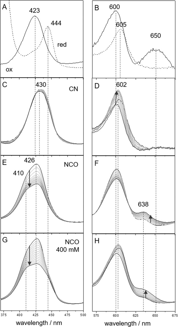
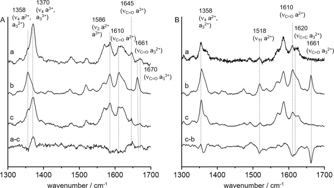
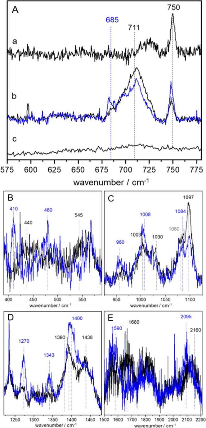
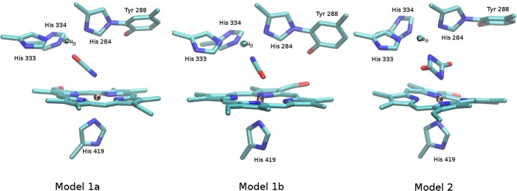
Heme-copper oxygen reductases are terminal respiratory enzymes, catalyzing the reduction of dioxygen to water and the translocation of protons across the membrane. Oxygen consumption is inhibited by various substances. Here we tested the relatively unknown inhibition of cytochrome c oxidase (CcO) with isocyanate. In contrast to other more common inhibitors like cyanide, inhibition with cyanate was accompanied with the rise of a metal to ligand charge transfer (MLCT) band around 638 nm. Increasing the cyanate concentration furthermore caused selective reduction of heme a. The presence of the CT band allowed for the first time to directly monitor the nature of the ligand via surface-enhanced resonance Raman (SERR) spectroscopy. Analysis of isotope sensitive SERR spectra in comparison with Density Functional Theory (DFT) calculations identified not only the cyanate monomer as an inhibiting ligand but suggested also presence of an uretdion ligand formed upon dimerization of two cyanate ions. It is therefore proposed that under high cyanate concentrations the catalytic site of CcO promotes cyanate dimerization. The two excess electrons that are supplied from the uretdion ligand lead to the observed physiologically inverse electron transfer from heme a3 to heme a.


Conflict of interest statement

The authors declare no competing interests.

Figures

Figure 1
Figure 1
CcO UV/Vis spectra in the Soret and α band in the (A,B) oxidized (straight) and reduced (dashed) state, after addition of (C,D) 200 mM CN, (E,F) 200 mM NCO and (G,H) 400 mM NCO. The spectra in (C–H) were recorded over a time span of 14 h. The arrows indicate the direction of incubation time.
Figure 2
Figure 2
SERR spectra at 413 nm (A) and 442 nm (B) excitation of immobilized CcO in (a) Phosphate buffer solution (PBS), (b) PBS + dithionite and (c) PBS subsequent to NCO incubation. The traces a-c and c-b correspond to the respective difference spectra.
Figure 3
Figure 3
(A) SERR spectra recorded with 647 nm excitation before (trace a) and after (trace b) immersion in KOCN (black line) and KO13C15N (blue line) buffer solution. Trace c shows the “slow” form of CcO in after immersion in KOCN buffer solution. (B–E) Same conditions as for A trace b but in an extended frequency range.
Figure 4
Figure 4
Structural models of the catalytic binuclear center harbouring single cyanate ions (model 1a, model 1b) or an uretdione ligand (model 2). For clarity, hydrogen atoms have been omitted in the representation.

References

    1. Aoyama H, et al. A peroxide bridge between Fe and Cu ions in the O2 reduction site of fully oxidized cytochrome c oxidase could suppress the proton pump. PNAS. 2009;106:2165–2169. doi: 10.1073/pnas.0806391106. - DOI - PMC - PubMed
    1. Pinakoulaki E, Vamvouka M, Varotsis C. Resonance Raman detection of the Fe2+−C−N modes in heme−copper oxidases: a probe of the active site. Inorg. Chem. 2004;43:4907–4910. doi: 10.1021/ic035216r. - DOI - PubMed
    1. Gardner MT, et al. Vibrational analysis of a molecular heme−copper assembly with a nearly linear FeIII–CN–CuII Bridge: insight into cyanide binding to fully oxidized cytochrome c oxidase. Inorg. Chem. 1996;35:6878–6884. doi: 10.1021/ic960575q. - DOI - PubMed
    1. Yano N, et al. X-ray structure of cyanide-bound bovine heart cytochrome c oxidase in the fully oxidized state at 2.0 Å resolution. Acta Cryst. F. 2015;71:726–730. doi: 10.1107/S2053230X15007025. - DOI - PMC - PubMed
    1. Thomson AJ, et al. The structure of the cytochrome a3-CuB site of mammalian cytochrome c oxidase as probed by MCD and EPR spectroscopy. J. Inorg. Biochem. 1985;23:187–197. doi: 10.1016/0162-0134(85)85025-X. - DOI - PubMed

Publication types

MeSH terms