. 2020 Mar 2;10(1):3788.
doi: 10.1038/s41598-020-60689-w.

A dynamic perfusion based blood-brain barrier model for cytotoxicity testing and drug permeation

Affiliations

A dynamic perfusion based blood-brain barrier model for cytotoxicity testing and drug permeation

Basma Elbakary et al. Sci Rep. .

Abstract

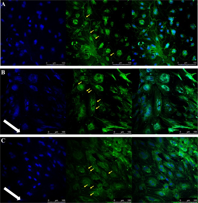
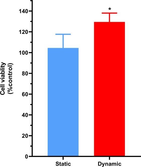
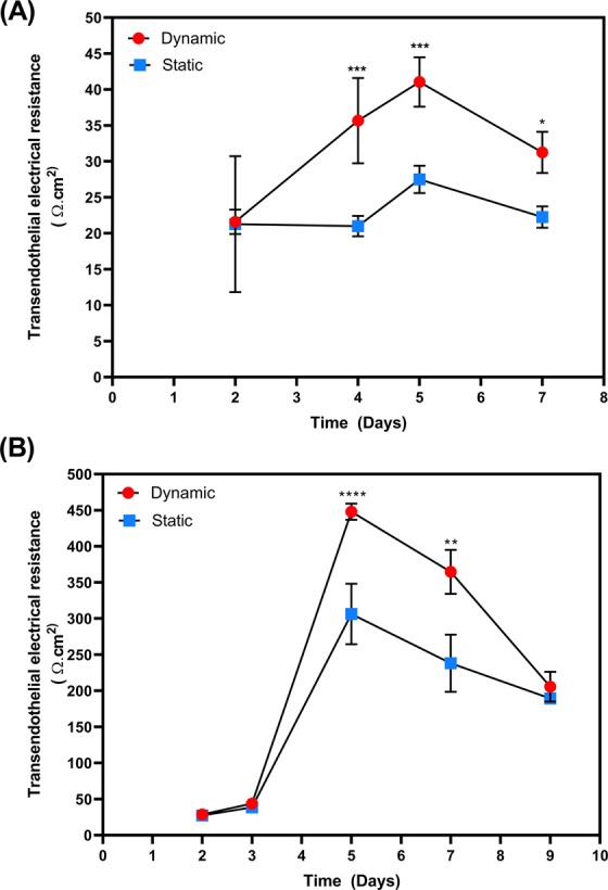
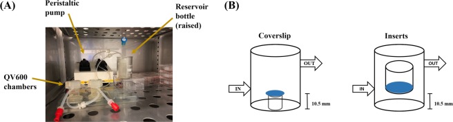
The blood-brain barrier (BBB) serves to protect and regulate the CNS microenvironment. The development of an in-vitro mimic of the BBB requires recapitulating the correct phenotype of the in-vivo BBB, particularly for drug permeation studies. However the majority of widely used BBB models demonstrate low transendothelial electrical resistance (TEER) and poor BBB phenotype. The application of shear stress is known to enhance tight junction formation and hence improve the barrier function. We utilised a high TEER primary porcine brain microvascular endothelial cell (PBMEC) culture to assess the impact of shear stress on barrier formation using the Kirkstall QuasiVivo 600 (QV600) multi-chamber perfusion system. The application of shear stress resulted in a reorientation and enhancement of tight junction formation on both coverslip and permeable inserts, in addition to enhancing and maintaining TEER for longer, when compared to static conditions. Furthermore, the functional consequences of this was demonstrated with the reduction in flux of mitoxantrone across PBMEC monolayers. The QV600 perfusion system may service as a viable tool to enhance and maintain the high TEER PBMEC system for use in in-vitro BBB models.


Conflict of interest statement

The authors declare no competing interests.

Figures

Figure 1
Figure 1
Immunocytochemistry images obtained following PBMEC staining for DAPI (left panel), ZO-1 (middle panel) and merged (right panel) when grown on bovine collagen (50 µg/mL) and fibronectin (7.5 µg/mL) coated coverslips. (A) PBMEC under static media conditions, (B) PBMEC grown under low flow (275 µL/min) and (C) PBMEC under high flow (550 µL/min) for 48 hours using the QV600. Images were taken using a Leica SP5 TCS II MP confocal microscope. White arrow indicates the direction of flow. Yellow arrows indicate formation of tight junctions.
Figure 2
Figure 2
Cellular viability of PBMEC grown on coverslips under static media and dynamic high flow (550 µL/min) for 96 hours, using an MTT assay. Dynamic results were normalised to the mean of the static results. n = 9 coverslips in 3 independent experiment. *p ≤ 0.05.
Figure 3
Figure 3
TEER measured following growth of PBMEC on permeable cell culture inserts (24-well, 0.33 cm2) under static and dynamic (550 µL/min) conditions. TEER of PBMEC were measured when grown on permeable inserts in the (A) absence and (B) presence of barrier forming additives. *p ≤ 0.05, **p ≤ 0.01, ***p ≤ 0.001 and ****p ≤ 0.0001. n = 12 in replicates of 3 in 4 independent experiments.
Figure 4
Figure 4
Immunocytochemisty images obtained following PBMEC staining for DAPI (left panel), ZO-1 (middle panel) and merged (right panel) when grown on permeable inserts. (A) PBMEC under static media conditions, (B) PBMEC grown under high flow (550 µL/min) for 24 hours and (C) PBMEC grown under high flow (550 µL/min) for 48 hours using the QV600. Images were taken using a Leica SP5 TCS II MP confocal microscope. White arrow indicates the direction of flow; yellow arrows indicate disrupted tight junction.
Figure 5
Figure 5
ZO-1 junctional fluorescence intensity and solidity as quantified by junctional regions and when normalised to static controls. *p ≤ 0.05.
Figure 6
Figure 6
Mitoxantrone flux across PBMEC grown on permeable inserts. Mitoxantrone transport in apical-to-basolateral (AB) (circles) or basolateral-to-apical (BA) (squares) under (A) static media or (B) when exposed to high flow (550 µL/min) for 48 hours with associated apparent membrane permeability (Papp) values in the AB or BA directions (C). *p ≤ 0.05, **p ≤ 0.01, ***p ≤ 0.001 and ****p ≤ 0.0001. n = 16 for static and dynamic, in 4 independent experiments.
Figure 7
Figure 7
A diagrammatical representation of: (A) the setup of the QV600 and (B) the location of coverslips in comparison to the use of permeable inserts.

References

    1. Daniels BP, et al. Immortalized human cerebral microvascular endothelial cells maintain the properties of primary cells in an in vitro model of immune migration across the blood brain barrier. Journal of neuroscience methods. 2013;212:173–179. doi: 10.1016/j.jneumeth.2012.10.001. - DOI - PMC - PubMed
    1. Abbott, N. J., Dolman, D. E., Drndarski, S. & Fredriksson, S. M. In Astrocytes 415–430 (Springer, 2012). - PubMed
    1. Lippmann ES, Al-Ahmad A, Azarin SM, Palecek SP, Shusta EV. A retinoic acid-enhanced, multicellular human blood-brain barrier model derived from stem cell sources. Sci. Rep. 2014;4:4160. doi: 10.1038/srep04160. - DOI - PMC - PubMed
    1. Patabendige A, Skinner RA, Abbott NJ. Establishment of a simplified in vitro porcine blood–brain barrier model with high transendothelial electrical resistance. Brain research. 2013;1521:1–15. doi: 10.1016/j.brainres.2012.06.057. - DOI - PMC - PubMed
    1. Cantrill CA, Skinner RA, Rothwell NJ, Penny JI. An immortalised astrocyte cell line maintains the in vivo phenotype of a primary porcine in vitro blood-brain barrier model. Brain research. 2012;1479:17–30. doi: 10.1016/j.brainres.2012.08.031. - DOI - PubMed