

Growth factors-based therapeutic strategies and their underlying signaling mechanisms for peripheral nerve regeneration
- PMID: 32123299
- PMCID: PMC7608263
- DOI: 10.1038/s41401-019-0338-1
Growth factors-based therapeutic strategies and their underlying signaling mechanisms for peripheral nerve regeneration
Abstract
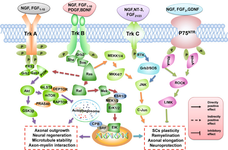
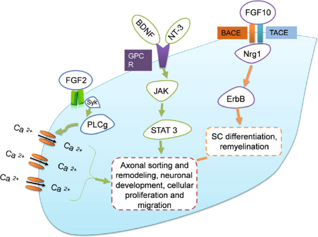
Peripheral nerve injury (PNI), one of the most common concerns following trauma, can result in a significant loss of sensory or motor function. Restoration of the injured nerves requires a complex cellular and molecular response to rebuild the functional axons so that they can accurately connect with their original targets. However, there is no optimized therapy for complete recovery after PNI. Supplementation with exogenous growth factors (GFs) is an emerging and versatile therapeutic strategy for promoting nerve regeneration and functional recovery. GFs activate the downstream targets of various signaling cascades through binding with their corresponding receptors to exert their multiple effects on neurorestoration and tissue regeneration. However, the simple administration of GFs is insufficient for reconstructing PNI due to their short half‑life and rapid deactivation in body fluids. To overcome these shortcomings, several nerve conduits derived from biological tissue or synthetic materials have been developed. Their good biocompatibility and biofunctionality made them a suitable vehicle for the delivery of multiple GFs to support peripheral nerve regeneration. After repairing nerve defects, the controlled release of GFs from the conduit structures is able to continuously improve axonal regeneration and functional outcome. Thus, therapies with growth factor (GF) delivery systems have received increasing attention in recent years. Here, we mainly review the therapeutic capacity of GFs and their incorporation into nerve guides for repairing PNI. In addition, the possible receptors and signaling mechanisms of the GF family exerting their biological effects are also emphasized.
Keywords: axonal regeneration; basic fibroblast growth factor; growth factors; nerve conduits; nerve growth factor; peripheral nerve injury; signaling cascade.
Conflict of interest statement
The authors declare no competing interests.
Figures

References
-
- Gu X, Ding F, Yang Y, Liu J. Construction of tissue engineered nerve grafts and their application in peripheral nerve regeneration. Prog Neurobiol. 2011;93:204–30. - PubMed
-
- Ichihara S, Inada Y, Nakamura T. Artificial nerve tubes and their application for repair of peripheral nerve injury: an update of current concepts. Injury. 2008;39(Suppl 4):29. - PubMed
-
- Sunderland S. A classification of peripheral nerve injuries producing loss of function. Brain. 1951;74:491–516. - PubMed
-
- Robinson LR. Traumatic injury to peripheral nerves. Muscle Nerve. 2000;23:863–73. - PubMed
Publication types
MeSH terms
Substances
LinkOut - more resources
Full Text Sources
Other Literature Sources
Medical
