

Spinal intramedullary epidermoid cysts: Three case presentations and literature review
- PMID: 32123605
- PMCID: PMC7049888
- DOI: 10.25259/SNI_540_2019
Spinal intramedullary epidermoid cysts: Three case presentations and literature review
Abstract
Background: True intramedullary epidermoid cysts (IECs) not associated with congenital anomalies or previous spinal procedures are extremely rare. In a review of the literature since 1992, only 29 such cases have been reported. Here, we add three new cases in this category.
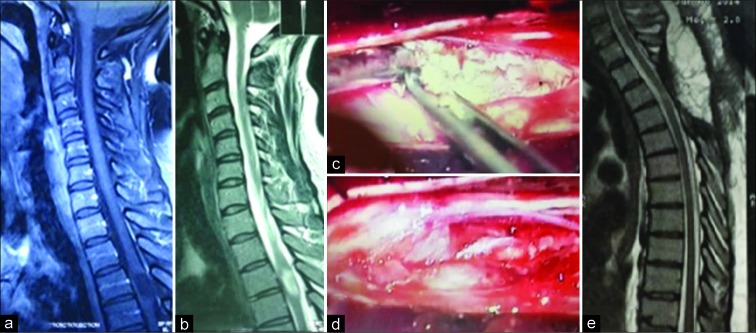
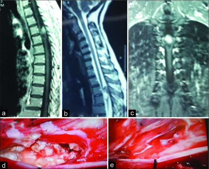
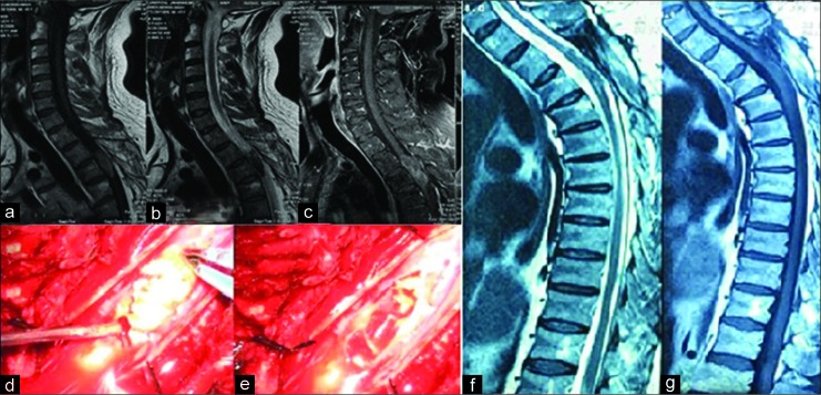
Case description: Three adults presented with spastic paraparesis attributed to thoracic IECs. Gross total microsurgical removal was achieved in two cases, while one case was a partial resection due to capsular adherence to the cord. In all three cases, patients sustained complete recoveries of neurological function and remained symptom free for an average of 5 years follow-up.
Conclusion: IECs are rare lesions; here, the three located in the thoracic spine, contributed to slow, progressive spastic paraparesis with/without incontinence, and resolved following total (2 patients) and partial (1 patient) resection.
Keywords: Epidermoid cyst; Intramedullary tumor; Paraparesis; Spinal cord; Thoracic spine.
Copyright: © 2020 Surgical Neurology International.
Conflict of interest statement
There are no conflicts of interest.
Figures
Similar articles
-
Adult intramedullary epidermoid cyst without spinal dysraphism: A case report.Surg Neurol Int. 2018 Jun 18;9:122. doi: 10.4103/sni.sni_117_18. eCollection 2018. Surg Neurol Int. 2018. PMID: 30009086 Free PMC article.
-
Intramedullary Spinal Epidermoid Cyst-A Rare Cause of Spastic Paraparesis.Asian J Neurosurg. 2024 Jun 4;19(2):309-311. doi: 10.1055/s-0044-1787049. eCollection 2024 Jun. Asian J Neurosurg. 2024. PMID: 38974452 Free PMC article.
-
Intramedullary epidermoid cysts in adults: Case report and updated literature review.Neurochirurgie. 2017 May;63(2):99-102. doi: 10.1016/j.neuchi.2017.01.004. Epub 2017 May 8. Neurochirurgie. 2017. PMID: 28495229 Review.
-
Isolated thoracic (D5) intramedullary epidermoid cyst without spinal dysraphism: A rare case report.J Pediatr Neurosci. 2015 Apr-Jun;10(2):133-6. doi: 10.4103/1817-1745.159206. J Pediatr Neurosci. 2015. PMID: 26167216 Free PMC article.
-
Spinal Intramedullary Epidermoid Cyst: Case Report and Updated Literature Review.World Neurosurg. 2020 Jul;139:39-50. doi: 10.1016/j.wneu.2020.03.207. Epub 2020 Apr 13. World Neurosurg. 2020. PMID: 32298825 Review.
Cited by
-
Intradural intramedullary epidermoid cyst in a 17-year-old male: An exceptionally rare case report and review of the literature.Int J Surg Case Rep. 2024 Mar;116:109331. doi: 10.1016/j.ijscr.2024.109331. Epub 2024 Feb 2. Int J Surg Case Rep. 2024. PMID: 38340621 Free PMC article.
-
Intramedullary mature teratoma with an exophytic component in an adult: Report of a case and literature review.Surg Neurol Int. 2020 Jul 11;11:187. doi: 10.25259/SNI_325_2020. eCollection 2020. Surg Neurol Int. 2020. PMID: 35592009 Free PMC article.
-
Discrepancy between MRI and intraoperative findings in a rare intramedullary epidermoid cyst: A case report and literature review.Radiol Case Rep. 2025 May 15;20(8):3755-3760. doi: 10.1016/j.radcr.2025.04.049. eCollection 2025 Aug. Radiol Case Rep. 2025. PMID: 40486160 Free PMC article.
-
Filum terminale infected epidermoid cysts in pediatric age group; A case series.World Neurosurg X. 2024 Sep 21;24:100408. doi: 10.1016/j.wnsx.2024.100408. eCollection 2024 Oct. World Neurosurg X. 2024. PMID: 39391069 Free PMC article.
-
Neurologic pathologies of the vertebral spine.Skeletal Radiol. 2024 Mar;53(3):419-436. doi: 10.1007/s00256-023-04428-y. Epub 2023 Aug 17. Skeletal Radiol. 2024. PMID: 37589755 Review.
References
-
- Agrawal M, Gour SS, Borkar SA. Unusual calcification in intramedullary epidermoid cyst. World Neurosurg. 2019;126:99–100. - PubMed
-
- Alvord EC., Jr Growth rates of epidermoid tumors. Ann Neurol. 1977;2:367–70. - PubMed
-
- Amato VG, Assietti R, Arienta C. Intramedullary epidermoid cyst: Preoperative diagnosis and surgical management after MRI introduction. Case report and updating of the literature. J Neurosurg Sci. 2002;46:122–6. - PubMed
-
- Babayev R, Abbasov B, Ekşi MŞ. Thoracic intramedullary epidermoid cyst-timely fashion diagnosis and treatment. Childs Nerv Syst. 2015;31:793–6. - PubMed
Publication types
LinkOut - more resources
Full Text Sources