Case Reports
. 2020 Jun;14(2):311-320.
doi: 10.1007/s12105-020-01140-4. Epub 2020 Mar 2.

Salivary Gland Cancer in the Era of Routine Next-Generation Sequencing

Affiliations
Case Reports

Salivary Gland Cancer in the Era of Routine Next-Generation Sequencing

Emilija Todorovic et al. Head Neck Pathol. 2020 Jun.

Abstract

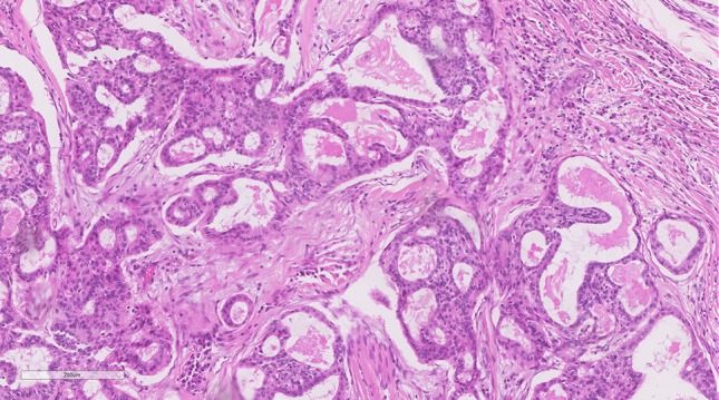
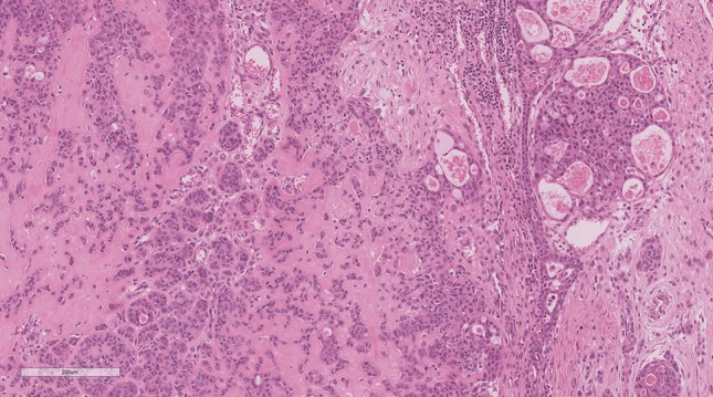
Next-Generation Sequencing (NGS) is being utilized with increasing frequency in the characterization of salivary gland tumours. The potential scenarios which may be encountered by using this technique in routine practice will be outlined in further text by drawing from our own clinical experience. These include oncocytic mucoepidermoid carcinomas with unusual variant morphology (and negative MAML2 fluorescent in-situ hybridization results), a diagnosis of ameloblastoma changed to adenoid cystic carcinoma (due to MYBL1 fusion presence), a salivary duct carcinoma with an ETV6-NTRK3 fusion (otherwise seen in secretory carcinomas) and novel fusion partners such as EWSR1-BEND2 (otherwise seen in pancreatic neuroendocrine carcinomas). As NGS continues to develop and more widespread clinical implementation increases, we must be cognisant of the need for proper interpretation and in some cases verification using a secondary technique, the limitations of this technique, and the ethical dilemmas one faces when encountering a novel fusion.

Keywords: Fluorescence; Fusions; In-situ hybridization; Next-generation sequencing; Salivary gland neoplasms.


Conflict of interest statement

No conflict of interest to disclose.

Figures

Fig. 1
Fig. 1
Oncocytic mucoepidermoid carcinoma with CRTC1-MAML2 fusion: Despite a negative MAML2 FISH result and relative lack of mucin, this tumour with predominantly solid nests of eosinophilic cells with low-grade nuclear features and focal ductular structures, had the appearance and staining pattern of an oncocytic mucoepidermoid carcinoma and proven as such by NGS with a CRTC1-MAML2 fusion
Fig. 2
Fig. 2
Adenoid cystic carcinoma with squamous differentiation and variant MYBL1-NFIB fusion: a Cystic spaces lined by basaloid cells with small hyperchromatic nuclei and peripheral palisading showing cribriform growth. b Focal squamous pearls are noted in an otherwise completely basaloid appearing neoplasm on the initial biopsy. This tumour originally called ameloblastoma in the nasal cavity was diagnosed as adenoid cystic carcinoma on the basis of an MYBL1-NFIB fusion by NGS
Fig. 3
Fig. 3
Adenoid cystic carcinoma with canalicular architecture and variant MYBL1-NFIB fusion: Well-demarcated islands formed by small basaloid cells with a combination of patterns including trabecular and canalicular growth. This palatal tumour was also diagnosed as adenoid cystic carcinoma on the basis of an MYBL1-NFIB fusion by NGS
Fig. 4
Fig. 4
Salivary duct carcinoma with ETV6-NTRK3 fusion: a Infiltrative nests of apocrine-appearing cells with high-grade cytologic atypia and extensive comedo-necrosis. b Diffuse AR and BRST-2 were seen (AR shown here). c The case showed no evidence of secretory carcinoma or low-grade morphology and was S100 negative (shown here). d. FISH showed an ETV6 rearrangement (shown here) confirming the initial incidental finding of ETV6-NTRK3 by NGS
Fig. 5
Fig. 5
Adenocarcinoma NOS with CRTC1-MAML2 fusion: Infiltrative glands with low to intermediate-grade cytologic features and lacking any morphologic, histochemical or immunohistochemical evidence of the squamous or mucinous features typical of mucoepidermoid carcinoma. However, the case was eventually shown to be CRTC1-MAML2 positive by NGS. While it may not be appropriate to change the diagnosis it is likely this tumour is related to, or is in fact a variant morphology in mucoepidermoid carcinoma
Fig. 6
Fig. 6
Adenocarcinoma NOS with a novel EWSR1-BEND2 fusion: Cribriform structures and individually infiltrative cells with high-grade nuclei and a signet-ring/lobular breast-like appearance in the dyscohesive cells. This tumour shows an unrecognizable morphology and was shown to have a novel EWSR1-BEND2 fusion by NGS. This type of finding may suggest a novel “cribriform and lobular adenocarcinoma” entity, but it is not appropriate to report clinically as it has no diagnostic or therapeutic implications at this time

References

    1. World Health Organization classification of tumours: pathology and genetics of head and neck tumours. 4th Edition [press release]. Lyon2017.
    1. Foote FW, Jr, Frazell EL. Tumors of the major salivary glands. Cancer. 1953;6(6):1065–1133. - PubMed
    1. Skalova A, Stenman G, Simpson RHW, Hellquist H, Slouka D, Svoboda T, et al. The role of molecular testing in the differential diagnosis of salivary gland carcinomas. Am J Surg Pathol. 2018;42(2):e11–e27. - PubMed
    1. Griffith CC, Schmitt AC, Little JL, Magliocca KR. New developments in salivary gland pathology: clinically useful ancillary testing and new potentially targetable molecular alterations. Arch Pathol Lab Med. 2017;141(3):381–395. - PubMed
    1. Dickson BC, Swanson D. Targeted RNA sequencing: A routine ancillary technique in the diagnosis of bone and soft tissue neoplasms. Genes Chromosomes Cancer. 2019;58(2):75–87. - PubMed

Publication types

MeSH terms