. 2020 Mar;56(3):772-782.
doi: 10.3892/ijo.2020.4968. Epub 2020 Jan 22.

Diallyl disulfide induces downregulation and inactivation of cofilin 1 differentiation via the Rac1/ROCK1/LIMK1 pathway in leukemia cells

Affiliations

Diallyl disulfide induces downregulation and inactivation of cofilin 1 differentiation via the Rac1/ROCK1/LIMK1 pathway in leukemia cells

Hui Ling et al. Int J Oncol. 2020 Mar.

Abstract

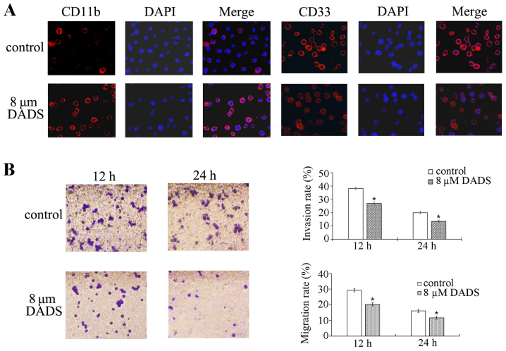
Cofilin is associated with cell differentiation; however, to the best of our knowledge, no data have indicated an association between the cofilin 1 pathway and leukemia cell differentiation. The present study investigated the involvement of the cofilin 1 signaling pathway in diallyl disulfide (DADS)‑induced differentiation and the inhibitory effects on the proliferation, migration, and invasion of human leukemia HL‑60 cells. First, it was identified that 8 µM DADS suppressed cell proliferation, migration and invasion, and induced differentiation based on the reduced nitroblue tetrazolium ability and increased CD11b and CD33 expression. DADS significantly downregulated the expression of cofilin 1 and phosphorylated cofilin 1 in HL‑60 leukemia cells. Second, it was verified that silencing cofilin 1 markedly promoted 8 µM DADS‑induced differentiation and the inhibitory effect on cell proliferation and invasion. Overexpression of cofilin 1 obviously suppressed 8 µM DADS‑induced differentiation and the inhibitory effect on cell proliferation and invasion. Third, the present study examined the mechanisms by which 8 µM DADS decreases cofilin 1 expression and activation. The results revealed that 8 µM DADS inhibited the mRNA and protein expression of Rac1, Rho‑associated protein kinase 1 (ROCK1) and LIM domain kinase 1 (LIMK1) as well as the phosphorylation of LIMK1 in HL‑60 cells, while 8 µM DADS enhanced the effects of the Rac1‑ROCK1‑LIMK1 pathway in cells overexpressing cofilin 1 compared with that in control HL‑60 cells. These results suggest that the anticancer function of DADS on HL‑60 leukemia cells is regulated by the Rac1‑ROCK1‑LIMK1‑cofilin 1 pathway, indicating that DADS could be a promising anti‑leukemia therapeutic compound.

Keywords: leukemia; diallyl disulfide; cofilin 1; differentiation; proliferation.


Figures

Figure 1
Figure 1
Effects of DADS on the differentiation induction, migration and invasion of the human leukemic cell line HL-60. (A) Expression of the cell surface differentiation markers CD11b and CD33 induced by DADS. Magnification, ×200. HL-60 cells were exposed to 8 µM DADS for 72 h and then fixed in 4% formaldehyde/PBS and permeabilized with 0.5% Triton X-100. CD11b and CD33 were detected by immunofluorescence (red, left panel). The DNA-intercalating dye DAPI was applied to recognize cell nuclei (blue, center panel). The right panel shows a merged image to highlight the nuclear pool of CD11b and CD33. (B) The effect of DADS on the migration and invasion of HL-60 cells. Magnification, ×200. HL-60 cells were treated with 8 µM DADS for 12 and 24 h. Cell migration and invasion were analyzed by Transwell migration and invasion assays, respectively. The migration rate is presented as the ratio of the mean migrated cell numbers between treated and untreated cells. The invasion rate is presented as the ratio of the mean cell numbers between treated and untreated cells. *P<0.05 vs. the control. DADS, diallyl disulfide.
Figure 2
Figure 2
Inhibitory effect of DADS on Rac1/ROCK1/LIMK1/cofilin 1 signaling in HL-60 cells. The cells were treated with 8 µM DADS for the indicated amounts of time. (A) Reverse transcription-semi-quantitative PCR was performed to determine the mRNA expression levels of cofilin 1, Rac1, ROCK1 and LIMK1. β-actin was used as an internal control for normalization. (B) Western blotting was performed to determine cofilin 1, p-cofilin 1, Rac1, ROCK1, LIMK1 and p-LIMK1 protein levels. β-actin was used as a loading control. The relative fold changes in mRNA or protein levels were compared with β-actin controls. The results are presented as the mean ± standard error of the mean from three independent experiments. *P<0.05 vs. the control (0 h group). DADS, diallyl disulfide; ROCK1, Rho-associated protein kinase 1; LIMK1, LIM domain kinase 1, p-, phosphorylated.
Figure 3
Figure 3
Effects of DADS and cofilin 1 miRNA expression plasmid transfection on the proliferation, differentiation and invasion of HL-60 cells. (A) Effect of DADS and cofilin 1 miRNA expression plasmid transfection on the viability of HL-60 cells. In total, 3×10⁴ cells were treated with 8 µM DADS for 48 h, and the viable cells were analyzed for cell proliferation. (B) Effect of DADS and cofilin 1 miRNA expression plasmid transfection on NBT reduction in HL-60 cells. HL-60 cells were treated with 8 µM DADS for 12, 24 and 48 h. The differentiation of HL-60 cells was identified by the decrease in NBT absorbance at 570 nm. $P<0.05 vs. the untreated HL-60 group; *P<0.05 vs. the untreated cofilin 1-miR group. (C) Effects of DADS and cofilin 1 miRNA expression plasmid transfection on the migration and invasion of HL-60 cells. Briefly, 8 µM DADS was added to HL-60 cells and cells transfected with vector or a cofilin 1 miRNA expression plasmid for 24 h. Cell migration and invasion were determined using Transwell migration and invasion assays, respectively. The migration rate is presented as the ratio of migrated cells between treated and untreated HL-60 cells. The invasion rate is presented as the ratio of the mean cell numbers between treated and untreated cells. *P<0.05 vs. the untreated cofilin 1-miR group. $P<0.05 vs. HL-60 cells (D) Expression of the cell surface differentiation markers CD11b and CD33 following DADS treatment and cofilin 1 miRNA expression plasmid transfection. Magnification, ×200. CD11b and CD33 were identified by immunofluorescence (red, left panel). The DNA-intercalating dye DAPI was used to identify cell nuclei (blue, center panel). The right panel displays a merged image to highlight the nuclear pool of CD11b and CD33. Data are presented at the mean ± standard error or the mean from three independent experiments. DADS, diallyl disulfide; miRNA, microRNA; NBT, nitroblue tetrazolium; OD, optical density; cofilin 1-miR, cells transfected with cofilin 1 miRNA.
Figure 4
Figure 4
Effects of DADS on the Rac1-ROCK1-LIMK1 signaling pathway in cofilin 1 gene-silenced HL-60 cells. Western blotting was used to detect Rac1, ROCK1, LIMK1 and cofilin 1 protein levels. β-actin served as a loading control. The relative fold changes in protein levels compared with the β-actin control were analyzed. 1, untreated HL-60 cells; 2, HL-60 cells treated with DADS; 3, vector/HL-60 cells; 4, vector/HL-60 cells treated with DADS; 5, cofilin 1-miR/HL-60 cells; and 6, cofilin 1-miR/HL-60 cells treated with DADS. Data are presented at the mean ± standard error or the mean from three independent experiments. $P<0.05 vs. the untreated HL-60 group; *P<0.05 vs. the untreated cofilin 1-silenced/HL-60 group. DADS, diallyl disulfide; ROCK1, Rho-associated protein kinase 1; LIMK1, LIM domain kinase 1; cofilin 1-miR, cells transfection with cofilin 1 miRNA overexpressing vector.
Figure 5
Figure 5
Effect of DADS on the proliferation, differentiation and invasion of cofilin 1-transfected HL-60 cells. (A) Effect of DADS on the viability of HL-60 cells, vector-transfected cells and cofilin 1-transfected cells. In total, 3×10⁴ cells were treated with 8 µM DADS for 48 h, and the viable cells were analyzed for cell proliferation. #P<0.01 vs. the untreated HL-60 group; &P<0.01 vs. the untreated vector group; $P<0.001 vs. the untreated HL-60 group and the untreated vector-transfected group; *P<0.01 vs. the untreated cofilin 1 group; @P<0.001 vs. the untransfected cells treated with DADS and the vector-transfected cells treated with DADS. (B) Effect of DADS on NBT reduction in HL-60 cells, vector-transfected cells and cofilin 1-transfected cells. HL-60 cells were treated with 8 µM DADS for 48 h. The differentiation of HL-60 cells was determined by the decrease in NBT absorbance at 570 nm. $P<0.05 vs. the untreated HL-60 group; &P<0.05 vs. the HL-60 group with DADS treatment; **P<0.01 vs. the vector group with DADS treatment. (C) Effects of DADS on the migration and invasion of HL-60 cells. Briefly, 8 µM DADS was added to HL-60 cells, vector-transfected cells and cofilin 1-transfected cells for 24 h. Cell migration and invasion were examined using Transwell migration and invasion assays, respectively. The migration rate was calculated as the ratio of migrated cells between DADS-treated and untreated HL-60 cells. The invasion rate was estimated by the ratio of mean cell numbers between treated and untreated cells. $P<0.05, $$P<0.01 vs. the untreated HL-60 group; *P<0.05, **P<0.01 vs. the untreated cofilin 1 group. (D) Expression of the cell surface differentiation markers CD11b and CD33 after DADS treatment and cofilin 1 transfection. Magnification, ×200. CD11b and CD33 were identified by immunofluorescence (red, left panel). The DNA-intercalating dye DAPI was used to identify cell nuclei (blue, center panel). The right panel displays a merged image to highlight the nuclear pool of CD11b and CD33. DADS, diallyl disulfide; OD, optical density; NBT, nitroblue tetrazolium.
Figure 6
Figure 6
Effects of DADS on the Rac1-ROCK1-LIMK1 signaling pathway in cofilin 1-transfected HL-60 cells. (A) Reverse transcription-semi-quantitative PCR was used to determine the mRNA expression levels of Rac1, ROCK1, LIMK1 and cofilin 1. β-actin was used as an internal control for normalization. (B) Western blotting was employed to detect cofilin 1, p-cofilin 1, Rac1, ROCK1, LIMK1 and p-LIMK1 protein levels. β-actin was regarded as a loading control. The relative fold changes in mRNA or protein levels were compared with β-actin controls. M, Maker; 1, untreated HL-60 cells; 2, HL-60 cells treated with DADS; 3, vector/HL-60 cells; 4, vector/HL-60 cells treated with DADS; 5, cofilin 1/HL-60 cells; and 6, cofilin 1/HL-60 cells treated with DADS. Data are presented at the mean ± standard error or the mean from three independent experiments. *P<0.05 vs. the untreated HL-60 group. DADS, diallyl disulfide; ROCK1, Rho-associated protein kinase 1; LIMK1, LIM domain kinase 1; p-, phosphorylated.
Figure 7
Figure 7
Role of DADS in leukemia cells. DADS negatively regulates the Rac1-ROCK1-LIMK1 signaling pathway, decreases cofilin 1 expression and suppresses its phosphorylation, which leads to inhibition of proliferation, migration and invasion, and differentiation induction in leukemia HL-60 cells. DADS, diallyl disulfide; ROCK1, Rho-associated protein kinase 1; LIMK1, LIM domain kinase 1.

References

    1. Mahmud H, ter Elst A, Scherpen FJG, de Boer TM, Kampen KR, de Haas V, Guryev V, Peppelenbosch MM, Kornblau SM, de Bont ESJM. Peptide microarray of pediatric acute myeloid leukemia is related to relapse and reveals involvement of DNA damage response and repair. Oncotarget. 2019;10:4679–4690. - PMC - PubMed
    1. Surveillance, Epidemiology, and End Results (SEER) Program, SEER*Stat Database: Incidence - SEER 9 Regs Research Data, Nov 2017 Sub (1973-2015) - Linked To County Attributes - Total U.S., 1969-2016 Counties. National Cancer Institute, DCCPS, Surveillance Research Program; https://www.seer.cancer.gov released April 2018, based on the November 2017 submission.
    1. Yi L, Su Q. Molecular mechanisms for the anti-cancer effects of diallyl disulfide. Food Chem Toxicol. 2013;57:362–370. doi: 10.1016/j.fct.2013.04.001. - DOI - PubMed
    1. Zhao J, Huang WG, He J, Tan H, Liao QJ, Su Q. Diallyl disulfide suppresses growth of HL-60 cell through increasing histone acetylation and p21WAF1 expression in vivo and in vitro. Acta Pharmacol Sin. 2006;27:1459–1466. doi: 10.1111/j.1745-7254.2006.00433.x. - DOI - PubMed
    1. Ling H, He J, Tan H, Yi L, Liu F, Ji X, Wu Y, Hu H, Zeng X, Ai X, et al. Identification of potential targets for differentiation in human leukemia cells induced by diallyl disulfide. Int J Oncol. 2017;50:697–707. doi: 10.3892/ijo.2017.3839. - DOI - PubMed

MeSH terms