. 2020 Apr;45(4):1081-1090.
doi: 10.3892/ijmm.2020.4497. Epub 2020 Feb 13.

Pirfenidone attenuates homocysteine‑induced apoptosis by regulating the connexin 43 pathway in H9C2 cells

Affiliations

Pirfenidone attenuates homocysteine‑induced apoptosis by regulating the connexin 43 pathway in H9C2 cells

Kai Chen et al. Int J Mol Med. 2020 Apr.

Abstract

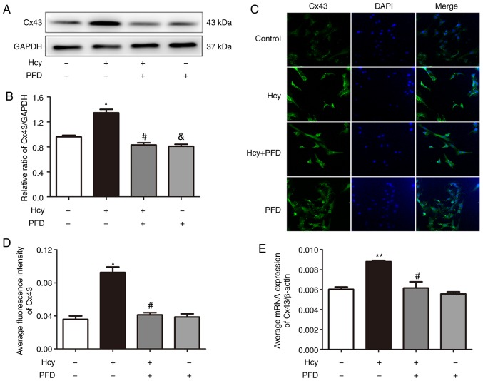
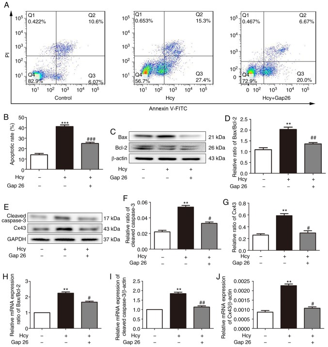
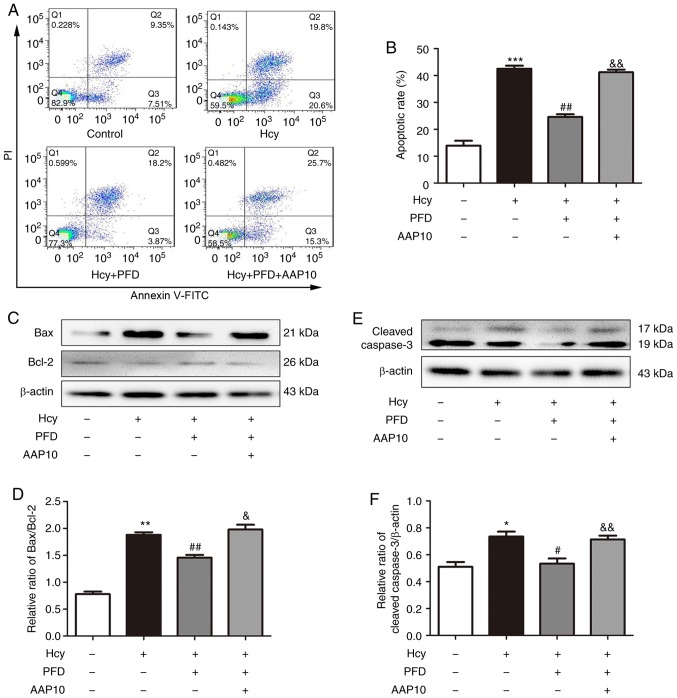
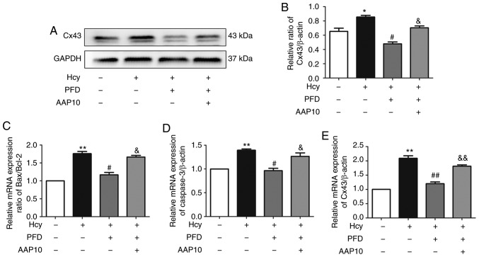
Pirfenidone (PFD) is an anti‑fibrotic agent that is clinically used in the treatment of idiopathic pulmonary fibrosis. PFD has been shown to exert protective effects against damage to orbital fibroblasts, endothelial cells, liver cells and renal proximal tubular cells; however, its effect on myocardial cell apoptosis remains unclear. The present study aimed to characterize the effects of PFD on homocysteine (Hcy)‑induced cardiomyocyte apoptosis and investigated the underlying mechanisms. H9C2 rat cardiomyocytes were pre‑treated with PFD for 30 min followed by Hcy exposure for 24 h. The effects of PFD on cell cytotoxicity were evaluated by CCK‑8 assay. The apoptosis rate of each group was determined by flow cytometry. The protein and mRNA levels of connexin 43 (Cx43), Bax, B‑cell lymphoma‑2 (Bcl‑2) and caspase‑3 were measured by western blot analysis and reverse transcription‑quantitative PCR, respectively. The present results demonstrated that the apoptotic rate increased following Hcy exposure, whereas the apoptotic rate significantly decreased following PFD pre‑treatment. Furthermore, the ratio of Bax/Bcl2 was upregulated following Hcy exposure, and Hcy upregulated the expression levels of cleaved caspase‑3 and Cx43. Notably, these effects were prevented by PFD. Additionally, the effects of PFD were inhibited by the Cx43 agonist, AAP10. In summary, the findings of the present study demonstrate that PFD protects H9C2 rat cardiomyocytes against Hcy‑induced apoptosis by modulating the Cx43 signaling pathway.


Figures

Figure 1
Figure 1
PFD prevents Hcy-induced cardiomyocyte cytotoxicity. (A) Dose-dependent cytotoxicity of Hcy towards H9c2 cells. H9c2 cells were exposed to 0-3 mmol/l Hcy for 24 h. (B) Cytotoxicity of PFD towards H9c2 cells. Cells were treated with PFD (0-1.5 mg/ml) for 24 h. (C) PFD alleviated Hcy-induced cytotoxicity in H9c2 cells. Cells were pre-treated with or without (0-1.5 mg/ml) PFD for 30 min and co-treated with 3 mM Hcy for 24 h. (D) The cellular morphology of H9C2 cardiomyocytes was examined under an inverted phase contrast microscope (magnification, ×40). Cell viability following treatment was detected by CCK-8 assay. *P<0.05, **P<0.01 vs. control; #P<0.05, ##P<0.01, ###P<0.001 vs. Hcy exposure. Data are shown as the means ± SE (n=3). Hcy, homocysteine; PFD, pirfenidone.
Figure 2
Figure 2
Effects of PFD on the Hcy-induced apoptosis, increased Bax/Bcl2 and cleaved caspase-3 expression levels in H9C2 cells. (A) PFD inhibited H9C2 cell apoptosis following Hcy-induced injury. PFD was used at 1 mg/ml. H9C2 cell apoptosis was determined using Annexin V/PI staining and flow cytometry analysis. (B) Statistical analysis of the apoptotic rate in each group. (C and E) The Hcy-induced increase in Bax/Bcl-2 and cleaved caspase-3 protein level was prevented by PFD. (D and F) Quantification of Bax/Bcl-2 and cleaved caspase-3. *P<0.05, **P<0.01, ***P<0.001 vs. control; #P<0.05, ##P<0.01 vs. Hcy treatment. Data are shown as the means ± SE (n=3). Hcy, homocysteine; PFD, pirfenidone.
Figure 3
Figure 3
(A, B and D) Fluorescence localization of the apoptotic proteins, Bax, Bcl-2 and caspase-3. Magnification, ×200. (C and E) Half of the relative fluorescence intensity of Bax/Bcl-2 and caspase-3 quantitative analysis. (F and G) Hcy-induced Bax/Bcl-2 and cleaved caspase-3 mRNA upregulation was prevented by PFD. *P<0.05 vs. control; #P<0.05, ##P<0.01 vs. Hcy treatment; &P<0.05 vs. Hcy treatment. Data are shown as the means ± SE (n=3). Hcy, homocysteine; PFD, pirfenidone.
Figure 4
Figure 4
Cx43 is involved in the Hcy-induced apoptosis of H9C2 cardiomyocytes. (A) Effects of Hcy and PFD on the expression of Cx43. (B) Statistical analysis of the expression of Cx43. (C) Fluorescence localization of the apoptosis protein Cx43. Magnification, ×200. (D) Half of the relative fluorescence intensity of Cx43 quantitative analysis. (E) The effect of PFD on Cx43 mRNA expression in H9C2 cells was determined by reverse transcription-quantitative PCR. *P<0.05, **P<0.01 vs. control; #P<0.05 vs. Hcy treatment; &P<0.05 vs. Hcy treatment. Data are shown as the means ± SE (n=3). Hcy, homocysteine; PFD, pirfenidone; Cx43, connexin 43.
Figure 5
Figure 5
(A) Pre-treatment with Gap26 inhibited H9C2 cell apoptosis following Hcy-induced injury. Gap26 was used at 0.5 µmol/l. (B) Statistical analysis of the rate of apoptosis in each group. (C and E) Hcy-induced upregulation of Bax/Bcl-2, cleaved caspase-3 and Cx43 protein levels was prevented by Gap26. (D, F and G) Quantification of Bax/Bcl-2, cleaved caspase-3 and Cx43. (H-J) Hcy-induced upregulation of Bax/Bcl-2, cleaved caspase-3 and Cx43 mRNA levels was prevented by Gap26. **P<0.01, ***P<0.001 vs. control; #P<0.05, ##P<0.01 vs. Hcy treatment. Data are shown as the means ± SE (n=3). Hcy, homocys-teine; PFD, pirfenidone; Cx43, connexin 43.
Figure 6
Figure 6
PFD protects H9C2 cardiomyocytes against Hcy-induced apoptosis via the Cx43 pathway. (A) Hcy-induced apoptosis was attenuated by PFD, and the effects of PFD were reversed by AAP10. AAP10 was used at 50 nmol/l. (B) Statistical analysis of the rate of apoptosis in each group. (C and E) Hcy-induced upregulation of Bax/Bcl-2 and cleaved caspase-3 protein level was prevented by PFD. The effects of PFD were prevented by AAP10. (D and F) Western blot analysis of Bax/Bcl-2 and cleaved caspase-3. *P<0.05, **P<0.01, ***P<0.001 vs. control; #P<0.05, ##P<0.01 vs. Hcy treatment; &P<0.05, &&P<0.01 vs. Hcy + PFD treatment. Data are shown as the means ± SE (n=3). Hcy, homocysteine; PFD, pirfenidone; Cx43, connexin 43.
Figure 7
Figure 7
(A) The Hcy-induced upregulation of Cx43 protein level was prevented by PFD. The effects of PFD were prevented by AAP10. (B) Western blot analysis of Cx43. (C-E) Hcy-induced upregulation of Bax/Bcl-2, cleaved caspase-3 and Cx43 mRNA levels was prevented by PFD. The effects of PFD were prevented by AAP10. *P<0.05, **P<0.01 vs. control; #P<0.05, ##P<0.01 vs. Hcy treatment; &P<0.05, &&P<0.01 vs. Hcy + PFD treatment. Data are shown as the means ± SE (n=3). Hcy, homocysteine; PFD, pirfenidone; Cx43, connexin 43.

References

    1. Shekhar A, Heeger P, Reutelingsperger C, Arbustini E, Narula N, Hofstra L, Bax JJ, Narula J. Targeted imaging for cell death in cardiovascular disorders. JACC Cardiovasc Imaging. 2018;11:476–493. doi: 10.1016/j.jcmg.2017.11.018. - DOI - PubMed
    1. Madonna R, Balistreri CR, De Rosa S, Muscoli S, Selvaggio S, Selvaggio G, Ferdinandy P, De Caterina R. Impact of sex differences and diabetes on coronary atherosclerosis and ischemic heart disease. J Clin Med. 2019;8 doi: 10.3390/jcm8010098. pii: E98. - DOI - PMC - PubMed
    1. Luo F, Das A, Chen J, Wu P, Li X, Fang Z. Metformin in patients with and without diabetes: A paradigm shift in cardiovascular disease management. Cardiovasc Diabetol. 2019;18:54. doi: 10.1186/s12933-019-0860-y. - DOI - PMC - PubMed
    1. Aminzadeh A, Mehrzadi S. Cardioprotective effect of levosimendan against homocysteine-induced mitochondrial stress and apoptotic cell death in H9C2. Biochem Biophys Res Commun. 2018;507:395–399. doi: 10.1016/j.bbrc.2018.11.049. - DOI - PubMed
    1. Fan CD, Sun JY, Fu XT, Hou YJ, Li Y, Yang MF, Fu XY, Sun BL. Astaxanthin attenuates homocysteine-induced cardiotoxicity in vitro and in vivo by inhibiting mitochondrial dysfunction and oxidative damage. Front Physiol. 2017;8:1041. doi: 10.3389/fphys.2017.01041. - DOI - PMC - PubMed