

Cutaneous microsporidiosis in an immunosuppressed patient
- PMID: 32125011
- PMCID: PMC8805087
- DOI: 10.1111/cup.13674
Cutaneous microsporidiosis in an immunosuppressed patient
Abstract
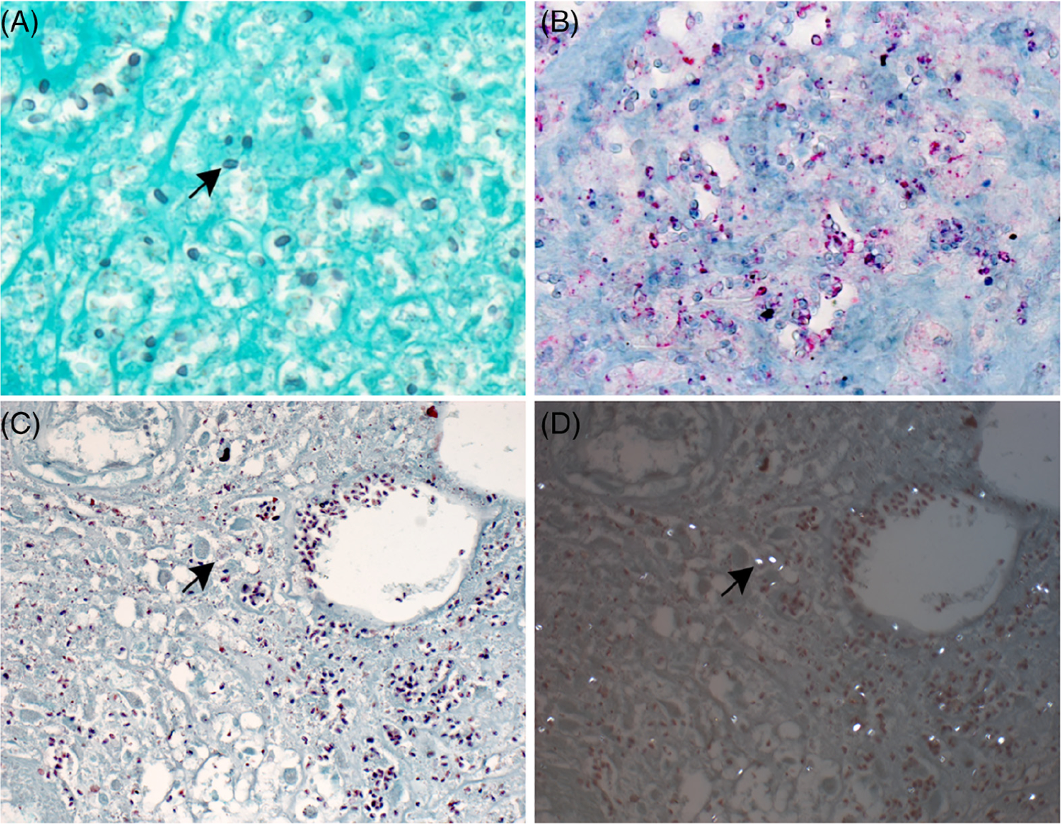
Microsporidia are a group of obligate intracellular parasites that naturally infect domestic and wild animals. Human microsporidiosis is an increasingly recognized multisystem opportunistic infection. The clinical manifestations are diverse with diarrhea being the most common presenting symptom. We present a 52-year-old woman with a history of amyopathic dermatomyositis complicated by interstitial lung disease managed with mycophenolate mofetil and hydroxychloroquine who presented with a 7-month history of recurrent subcutaneous nodules as well as intermittent diarrhea and chronic sinusitis. A punch biopsy showed superficial and deep lymphocytic and granulomatous dermatitis with focal necrosis. Tissue stains for microorganisms revealed oval 1 to 3 μm spores within the necrotic areas in multiple tissue stains. Additional studies at the Centers for Disease Control and Prevention confirmed cutaneous microsporidiosis. This case is one of very few confirmed examples of cutaneous microsporidiosis reported in the literature.
Keywords: cutaneous; immunosuppression; microsporidia; microsporidiosis.
© 2020 John Wiley & Sons A/S. Published by John Wiley & Sons Ltd.
Figures


References
-
- Field AS, Milner DA. Intestinal microsporidiosis. Clin Lab Med. 2015; 35(2):445–459. - PubMed
-
- Nagpal A, Pritt BS, Lorenz EC, et al. Disseminated microsporidiosis in a renal transplant recipient: case report and review of the literature. Transpl Infect Dis. 2013;15(5):526–532. - PubMed
-
- Kester KE, Turiansky GW, McEvoy PL. Nodular cutaneous microsporidiosis in a patient with AIDS and successful treatment with long-term clindamycin therapy. Ann Intern Med. 1998;128(11):911–914. - PubMed
