

ESHRD: deconvolution of brain homogenate RNA expression data to identify cell-type-specific alterations in Alzheimer's disease
- PMID: 32125278
- PMCID: PMC7093163
- DOI: 10.18632/aging.102840
ESHRD: deconvolution of brain homogenate RNA expression data to identify cell-type-specific alterations in Alzheimer's disease
Abstract
Objective: We describe herein a bioinformatics approach that leverages gene expression data from brain homogenates to derive cell-type specific differential expression results.
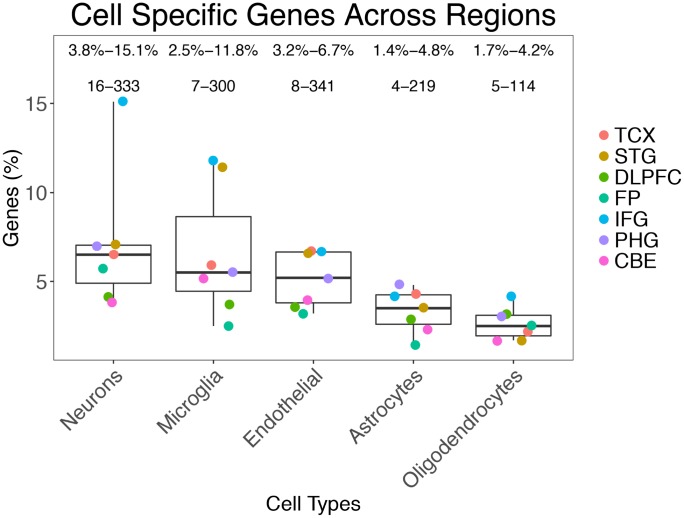
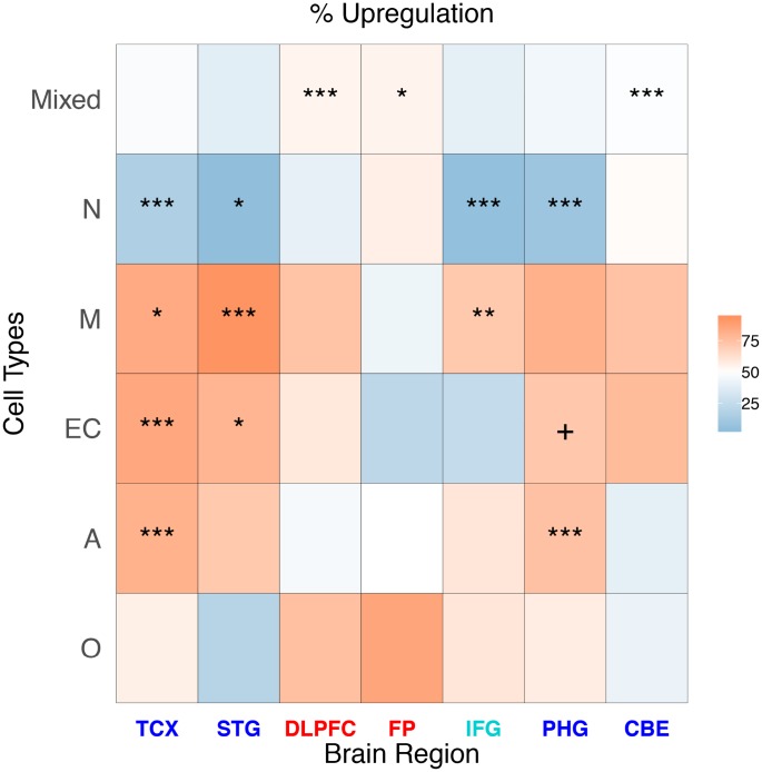
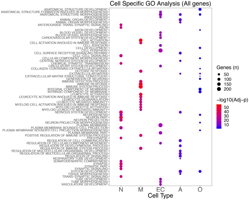
Results: We found that differentially expressed (DE) cell-specific genes were mostly identified as neuronal, microglial, or endothelial in origin. However, a large proportion (75.7%) was not attributable to specific cells due to the heterogeneity in expression among brain cell types. Neuronal DE genes were consistently downregulated and associated with synaptic and neuronal processes as described previously in the field thereby validating this approach. We detected several DE genes related to angiogenesis (endothelial cells) and proteoglycans (oligodendrocytes).
Conclusions: We present a cost- and time-effective method exploiting brain homogenate DE data to obtain insights about cell-specific expression. Using this approach we identify novel findings in AD in endothelial cells and oligodendrocytes that were previously not reported.
Methods: We derived an enrichment score for each gene using a publicly available RNA profiling database generated from seven different cell types isolated from mouse cerebral cortex. We then classified the differential expression results from 3 publicly accessible Late-Onset Alzheimer's disease (AD) studies including seven different brain regions.
Keywords: RNA sequencing; brain homogenates; endothelial cells; laser capture microdissection; oligodendrocytes.
Conflict of interest statement
Figures
References
-
- Mastroeni D, Nolz J, Sekar S, Delvaux E, Serrano G, Cuyugan L, Liang WS, Beach TG, Rogers J, Coleman PD. Laser-captured microglia in the Alzheimer’s and Parkinson’s brain reveal unique regional expression profiles and suggest a potential role for hepatitis B in the Alzheimer’s brain. Neurobiol Aging. 2018; 63:12–21. 10.1016/j.neurobiolaging.2017.10.019 - DOI - PMC - PubMed
-
- Sekar S, McDonald J, Cuyugan L, Aldrich J, Kurdoglu A, Adkins J, Serrano G, Beach TG, Craig DW, Valla J, Reiman EM, Liang WS. Alzheimer’s disease is associated with altered expression of genes involved in immune response and mitochondrial processes in astrocytes. Neurobiol Aging. 2015; 36:583–91. 10.1016/j.neurobiolaging.2014.09.027 - DOI - PMC - PubMed
-
- Liang WS, Dunckley T, Beach TG, Grover A, Mastroeni D, Ramsey K, Caselli RJ, Kukull WA, McKeel D, Morris JC, Hulette CM, Schmechel D, Reiman EM, et al.. Altered neuronal gene expression in brain regions differentially affected by Alzheimer’s disease: a reference data set. Physiol Genomics. 2008; 33:240–56. 10.1152/physiolgenomics.00242.2007 - DOI - PMC - PubMed
Publication types
MeSH terms
Grants and funding
- U01 AG046152/AG/NIA NIH HHS/United States
- P50 AG016574/AG/NIA NIH HHS/United States
- U01 AG046170/AG/NIA NIH HHS/United States
- R01 AG032990/AG/NIA NIH HHS/United States
- R01 NS080820/NS/NINDS NIH HHS/United States
- R21 NS093222/NS/NINDS NIH HHS/United States
- P01 AG017216/AG/NIA NIH HHS/United States
- R01 AG018023/AG/NIA NIH HHS/United States
- U01 AG006786/AG/NIA NIH HHS/United States
- R01 AG041232/AG/NIA NIH HHS/United States
- U01 AG046139/AG/NIA NIH HHS/United States
- P01 AG003949/AG/NIA NIH HHS/United States
- U24 NS072026/NS/NINDS NIH HHS/United States
- P30 AG019610/AG/NIA NIH HHS/United States
- P50 AG025711/AG/NIA NIH HHS/United States
LinkOut - more resources
Full Text Sources
Medical
