. 2020 Jun 1;130(6):3137-3150.
doi: 10.1172/JCI130426.

IRF4 instructs effector Treg differentiation and immune suppression in human cancer

Affiliations

IRF4 instructs effector Treg differentiation and immune suppression in human cancer

Giorgia Alvisi et al. J Clin Invest. .

Abstract

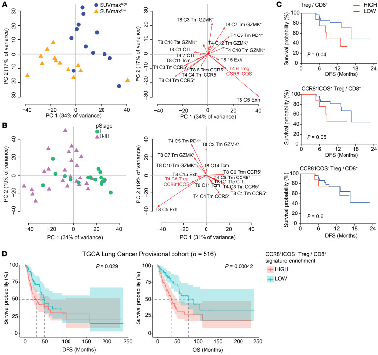
The molecular mechanisms responsible for the high immunosuppressive capacity of CD4+ Tregs in tumors are not well known. High-dimensional single-cell profiling of T cells from chemotherapy-naive individuals with non-small-cell lung cancer identified the transcription factor IRF4 as specifically expressed by a subset of intratumoral CD4+ effector Tregs with superior suppressive activity. In contrast to the IRF4- counterparts, IRF4+ Tregs expressed a vast array of suppressive molecules, and their presence correlated with multiple exhausted subpopulations of T cells. Integration of transcriptomic and epigenomic data revealed that IRF4, either alone or in combination with its partner BATF, directly controlled a molecular program responsible for immunosuppression in tumors. Accordingly, deletion of Irf4 exclusively in Tregs resulted in delayed tumor growth in mice while the abundance of IRF4+ Tregs correlated with poor prognosis in patients with multiple human cancers. Thus, a common mechanism underlies immunosuppression in the tumor microenvironment irrespective of the tumor type.

Keywords: Adaptive immunity; Cancer immunotherapy; Immunology; Oncology; T cells.


Conflict of interest statement

Conflict of interest: The Laboratory of Translational Immunology receives reagents in-kind from BD Biosciences Italy as part of a collaborative research agreement.

Figures

Figure 1
Figure 1. IRF4 identifies effector Tregs enriched in human tumors.
(A) UMAP analysis of concatenated CD4+ T cells (1,500 cells/sample) from tumor (n = 53), adjacent lung tissue (n = 45), and blood (n = 22) samples from patients with NSCLC. (B) UMAP of relative marker expression by concatenated CD4+ T cells from the same samples in A. (C) Manual gating analysis of CD4+CD25+FOXP3+ Tregs expressing IRF4+ by flow cytometry. Numbers indicate the percentage of positive cells. (D) Summary plot representing the IRF4 expression in CD4+ Tregs and conventional T (Tconv cells; defined as CD25loFoxP3) cells from the same patients as in A (***P < 0.0005, ****P < 0.0001, Kruskal-Wallis test). (E) Representative distribution by flow cytometry (top) and summary of the percentage of expression of selected markers (bottom) in tumor-infiltrating IRF4+ and IRF4 Tregs and Tconv cells (*P < 0.01, ***P < 0.0005, ****P < 0.0001, nonparametric Friedman test). (F) Box plot showing the log2(TPM + 1) expression of IRF4 transcript across 9 CD4+ T cells clusters as identified by single-cell RNA-seq (25). Each dot represents a single cell (*P ≤ 0.01, ****P ≤ 0.0001, Wilcoxon test). (G) t-SNE plots illustrating the expression of selected genes in single CD4+ T cells from lung tumor lesions. Cell clusters, depicted on the left, were identified as in F.
Figure 2
Figure 2. Transcriptional and functional profiling defines the effector and enhanced suppressive nature of IRF4+ Tregs.
(A) Representative CCR8 and ICOS expression in tumor-infiltrating CD25hiCD127lo Treg subsets defined by IRF4 expression and t percentage of IRF4 expression in tumor-infiltrating Tregs gated as CCR8ICOS or CCR8+ICOS+. (B) Heatmap of differentially expressed genes (DEGs) in the FACS-sorted CCR8+ICOS+ versus ICOSCCR8 tumor-infiltrating Tregs, as obtained by RNA-seq (FDR < 0.05). Selected DEGs are indicated. For some genes, protein names are indicated. (C) Hallmark gene sets (MsigDB; as obtained by GSEA) significantly enriched in cells sorted as in B. (D) Transcription factor binding motif (TFBM) enrichment analysis by pScan of RNA-seq data obtained as in B. Colored dots indicate significant hits. (E) CFSE-labeled CD4+ CD25 T (Tconv) cells dilution from a representative blood sample. Tconv cells were cocultured with Suppression Inspector MACSiBead beads and different ratios of intratumoral Treg subsets for 5 days. Data are representative of 5 independent experiments. (F) Tumor volumes in FoxP3EGFP-cre-ERT2(control) (n = 7) or Irf4fl/flFoxP3EGFP-cre-ERT2 (n = 5) mice following the administration of tamoxifen. Tumor curves in individual mice and mean ± SEM of the same cohort are shown. **P < 0.01, paired Student’s t test.
Figure 3
Figure 3. Irf4 and its partner Baft directly and indirectly control a molecular program of effector Treg differentiation and suppression.
(A) Identification of a shared gene expression signature between tumor-infiltrating human CCR8+ICOS+ Tregs versus CCR8ICOS Tregs and murine Tregs (36). (B) Venn diagram of the number of genes of the tumor-specific Treg signature obtained as in A that are differentially expressed in splenic Tregs from Batf–/– and Irf4–/– mice. Genes controlled only by Batf (n = 10) were of limited interest and thus not further investigated. (C) List of tumor-infiltrating Treg genes that are dependent on the expression of Irf4 or Irf4 and Batf. All genes are induced, except for Plac8, which is repressed (indicated in light blue). Those genes directly controlled by IRF4 binding to the genome, as obtained from ChIP-seq analysis of murine Tregs, are highlighted. Genomic binding of Irf4 to the DNA for selected genes is depicted.
Figure 4
Figure 4. Abundance of CCR8+ICOS+ intratumoral Tregs is associated with multiple features of T cell exhaustion.
(A) Experimental workflow. (B) Heatmaps of the relative expression, depicted as integrated MFI (iMFI: MFI × percentage of antigen expression) of markers (columns) in discrete CD4+ (left) and CD8+ (right) Phenograph clusters (rows). Tm, memory; Tcm, central memory; Tn, naive; Exh, exhausted; Act, activated; CTL, cytotoxic T lymphocyte. Tte, terminal effector. Data are further metaclustered to group subpopulations with similar immune-phenotypes. The median frequency of each PhenoGraph cluster in the different compartments is depicted by using Balloon plots. *P < 0.05; **P < 0.01; ***P < 0.001; ****P < 0.0001, tumor versus blood or versus normal tissue samples; 2-way ANOVA with Bonferroni’s post hoc test. (C) Correlogram showing Pearson correlation between frequencies of CD4+ (T4) and CD8+ (T8) PhenoGraph clusters in tumor samples from 45 patients with early-stage (I–III) non–small-cell lung cancer (NSCLC). Nonsignificant correlations (P value > 0.05) were left blank.
Figure 5
Figure 5. CCR8+ICOS+ Tregs define a signature of disease progression in NSCLC.
(A) Left: Principal component analysis (PCA) plot showing the distribution of patients (n = 48) according to the frequency of CD4+ and CD8+ PhenoGraph clusters in each patient (Tm, memory; Tcm, central memory). Patients were classified according to pathological stage (pStage) I, II, or III of the International TNM classification. Right: PCA loading plot of PhenoGraph clusters most contributing to the PCA output on the left. (B) Left: PCA plot showing the distribution of patients (n = 26) according to the frequency of CD4+ and CD8+ PhenoGraph clusters in each patient. The cohort was subdivided in 2 groups according to the median distribution of maximum standardized uptake value (SUVmax). Right: PCA loading plot as in A. (C) Kaplan-Meier progression-free survival curves according to the intratumoral frequencies of Tregs subsets over CD8+ T cells in each patient (n = 38). The cohort was subdivided in 2 groups according to the percentile rank (set at 0.8). The P value was calculated by Gehan Breslow-Wilcoxon test. (D) Kaplan-Meier disease-free survival (DFS) and overall survival (OS) curves in the TCGA lung adenocarcinoma (LUAD) lung cancer cohort (n = 516). Patients were grouped by percentile rank (set at 0.8) according to the enrichment of the CCR8+ICOS+ bulk Treg signature (as obtained in Figure 2B) as relative to the CD8+ T cell signature. + indicates censored observations. P values were calculated by multivariate Cox regression. Dotted lines indicate the time at which 50% of the cohort was still free of the event.

References

    1. Speiser DE, Ho PC, Verdeil G. Regulatory circuits of T cell function in cancer. Nat Rev Immunol. 2016;16(10):599–611. doi: 10.1038/nri.2016.80. - DOI - PubMed
    1. Tanaka A, Sakaguchi S. Regulatory T cells in cancer immunotherapy. Cell Res. 2017;27(1):109–118. doi: 10.1038/cr.2016.151. - DOI - PMC - PubMed
    1. Wing K, et al. CTLA-4 control over Foxp3+ regulatory T cell function. Science. 2008;322(5899):271–275. doi: 10.1126/science.1160062. - DOI - PubMed
    1. Larmonier N, et al. Tumor-derived CD4(+)CD25(+) regulatory T cell suppression of dendritic cell function involves TGF-beta and IL-10. Cancer Immunol Immunother. 2007;56(1):48–59. - PMC - PubMed
    1. Chen ML, et al. Regulatory T cells suppress tumor-specific CD8 T cell cytotoxicity through TGF-beta signals in vivo. Proc Natl Acad Sci USA. 2005;102(2):419–424. doi: 10.1073/pnas.0408197102. - DOI - PMC - PubMed

Publication types

Substances